Surgery, Gastroenterology and Oncology

|
|
Background: Cancer is considered as the second leading cause of death and the most popular kind of gastrointestinal cancer is colorectal cancer that resulted from genetic alterations progressed during a lifetime. In human cancer, the most frequently mutated oncogenes but have not produced to therapeutic attack are RAS genes (KRAS, NRAS and HRAS). Methotrexate is a cytotoxic chemotherapeutic agent, as it acts on cell cycle. Aims: This study aims to investigate the cytotoxicity effect MEX2R and MEX2O in human colorectal cells and to compare its action with MTX. MEX2R and MEX2O investigational agents as K-RAS oncogene blocker which inhibits the cell proliferation by targeting the cell cycle.
Methods: LST 174 colorectal cell line was grown in RPMI- 1640 media supplement with
10% heat inactivated fetal bovine serum and antibiotic 100IUs/ml penicillin with 100 mg/ml streptomycin. The cells were incubated with agents and drugs for 24 hours. Cells were inoculated in 96-well microtiter plate (105 cells/well) for 48 hrs. The final concentration of the solvent never exceeded 0.1%. Triplicates were prepared for each individual dose. Color intensity was measured in an ELISA reader. The viability and inhibition % of cancer cell line after the specified time were then detected and the concentration that inhibit 50% of cell viability (IC50) was fitted and calculated.
Results: There was no significant increase (P> 0.05) in cell growth inhibition as compared with control group at conc. (0.5,1,5 mg/ml) of MEX2R, while there was a highly significant increase (P£0.05) in inhibition of growth LST174 cells in conc. (25, 125,625 mg/ml). Additionally, the conc.of drug that needed to inhibit a biological process or response by 50% (IC50) is about 411 mg/ml . The result showed that MEX2O has significant difference (P£0.05) in LST174 cells growth inhibition in all concentrations (0.5,1.5.25,125,625 & 1000 mg/ml) when compared with control(untreated) group. While, the concentration of drug that needed to inhibit a biological process or response by 50% (IC50) is about 317.69 mg/ml. Also, the result showed that MTX has significant increase (P£0.05) in growth inhibition of LST174 cells in all concentrations (0.5,1,5,25,125,625,1000 mg/ml) when compared with control group. While, the results showed that the conc. of drug that needed to inhibit a biological process or response by 50% (IC50) is about 803.8 mg/ml.
Conclusion: MEX2R and MEX2O might be have a greater value in the treatment of this type of cancer than MTX as they act on the cell cycle as the tested drugs MEX2R & MEX2O acted on the cell cycle by targeting one of the main troubles that interfere with treatment of colorectal cancer which is K-RAS gene mutation and inhibit the cell proliferation.
Introduction
Cancer is considered as the second leading cause of death and a major public health problem after the cardiovascular diseases in worldwide (1). The most common type of gastrointestinal cancer is colorectal cancer (CRC), which results from genetic alterations that progress over a lifetime (2). According to GLOBOCAN 2018, the CRC incidence rate is approximately 10.2% of all new cases, and the mortality rate is approximately 9.2% of all cancer deaths.
One of the major factors that caused cancer is molecular genetic defects, many drugs were designed to reach at the affected site that block the cancer growth by interfering with particular molecules included in carcinogenesis and they may be more effective than conventional treatments and less harmful to normal cells, these are called targeted anti cancer therapy (3).
In human cancer, the most frequently mutated oncogenes that have not been produced for therapeutic attack are RAS genes (KRAS, NRAS, and HRAS), which are present in approximately 90% of pancreatic cancers, 45% of colon cancers, and 35% of lung cancers (4). The Kirsten rat sarcoma viral (KRAS) isoform mutation is predominant (approximately 86%), and mutations in exon 2 are most common among CRCs, with frequencies of 30 50%. While mutations of its exon 3 and 4 have lower effect, only show cause in about 1% and 4% (5).
In oncology, the most effective therapy used for the treatment of CRC is the anti-epidermal growth factor receptor (EGFR) antibody. However, KRAS mutations in patients with CRC are resistant to anti-EGFR therapy (6).
One of the most important targets for drug development is the KRAS signaling pathway due to these high occurrences either through direct K RAS targeting by binding to specific pockets on KRAS or inhibiting the GTPase activity of KRAS. These agents also have a similar strategy by recognizing the GDP analog SML 8 73 1 and the prodrug derivative of its SML 10 70 1, which blocks K RAS processing by covalently binding to G12C mutant K RAS, which is under investigation (7).
Other agents are prenyltransferase inhibitors (PTIs) that target RAS-membrane interactions and suppress their post-translational modifications (8). Salirasib (farnesylthiosalicylic acid) inhibits the farnesyltransferase (FTase) enzyme and suppresses proper plasma membrane attachment by interrupting the binding of persistently active RAS proteins to the plasma membrane; however, these agents do not exhibit clinical efficacy as single agents (9). Additionally, e GGTI 2417 and GGTI 241, 8 which t inhibit geranyl transferase (GGTI, do e not show e any clinical efficacy when used as a single agent or when combined with FTI, because they cause da toxicity s in clinical trials and l further clinical development (10).
On the other hand, the drugs that act by blocking RAF MAPK, and PI3 kinase downstream pathways are being examined in the clinic and they give different effects according to many factors like definite mutational variant and type of tissue (11).
Alternatively, inhibitor resistance has been attributed to many factors, such as negative feedback mechanisms, severe side effects, and reflexive activation of other downstream signaling partners of RAS (12 14).
Methotrexate (MTX) is a chemotherapeutic cyto-toxic agent that acts on the cell cycle (15). MTX limits the synthesis of thymidine and purine nucleotides and inhibits dihydrofolate reductase by interfering with DNA synthesis (16,17). This antifolate agent has severe adverse effects on the intestinal epithelium and bone marrow; therefore, there is a risk of hemorrhage that occurs spontaneously and is a life-threatening infection.
In this study, MEX2R and MEX2O, which are investigational agents, acted as KRAS oncogene blockers by inhibiting cell proliferation by targeting the cell cycle, and tested the cytotoxic effect in human colorectal cells.
Methods
Chemicals and Reagents
MEX2O and MEX2R (investigational agents), methotrexate injectable vial 50 mg / 5 ml (KOCAK FARMA), penicillin/streptomycin purchased from Sigma Aldrich (USA), MTT(3 (4,5 dimethyl 2 thiazolyl) 2,5 diphenyl 2H tetrazolium bromide) were purchased from Bioworld (USA), RPMI 1640 media (US Biological, USA) was supplemented with 10% heat-inactivated fetal bovine serum (Gibco, Germany), and all other agents were of analytical grade and used as received.
Cell Lines and Drugs Treatment
The LST 174 colorectal cell line was grown in RPMI 1640 medium supplemented with 10% heat-inactivated fetal bovine serum and antibiotic 100IUs/ml penicillin and 100 ?g/ml streptomycin. Cells were cultured at 37 °C in a 95% humidified atmosphere with 5% CO2. The cells were incubated with the agents and drugs for 24 h, and the attached cells were harvested for subsequent analysis.
In Vitro Cytotoxicity Assessment
The cytotoxic activity of the NCD was evaluated against the RMS cell line using an MTT assay. These cells were maintained in DMEM supplemented with 10% heat-inactivated fetal bovine serum (FBS). To maintain the cells in an exponential phase cellular suspension, aliquots were refed with fresh DMEM two or three times per week. The cells were inoculated in 96 well microtiter plate (105 cells/well) for 48 h. To allow the growth of the cell monolayer on the wall of the microtiter plate. An optimization protocol was developed for the toxicity screening of compounds in the cell monolayer. The test compounds were freshly dissolved in dimethyl sulfoxide (DMSO) and diluted in DMEM. The final solvent concentration did not exceed 0.1%. Samples were prepared in triplicate for each dose. Monolayer cells were incubated with the target compounds (NCD, DOX, MTX and MT1) for 24 h, at 37°C, 5% CO2 and incubated at 37°C in a humidified atmosphere. Color intensity was measured using an ELISA reader. The viability and inhibition % of the cancer cell line after the specified time were then determined, and the concentration that inhibit 50% of cell viability (IC50) was fitted and calculated.
Statistical Analysis
Statistical analyses were performed using IBM SPSS version 20. Because we compare more than 2 groups ANOVA test was used, means ±S.E used to express the data. Statistical significance was set at P < 0.05. Fifty percent inhibitory concentrations were determined by blotting inhibition% versus compound concentration.
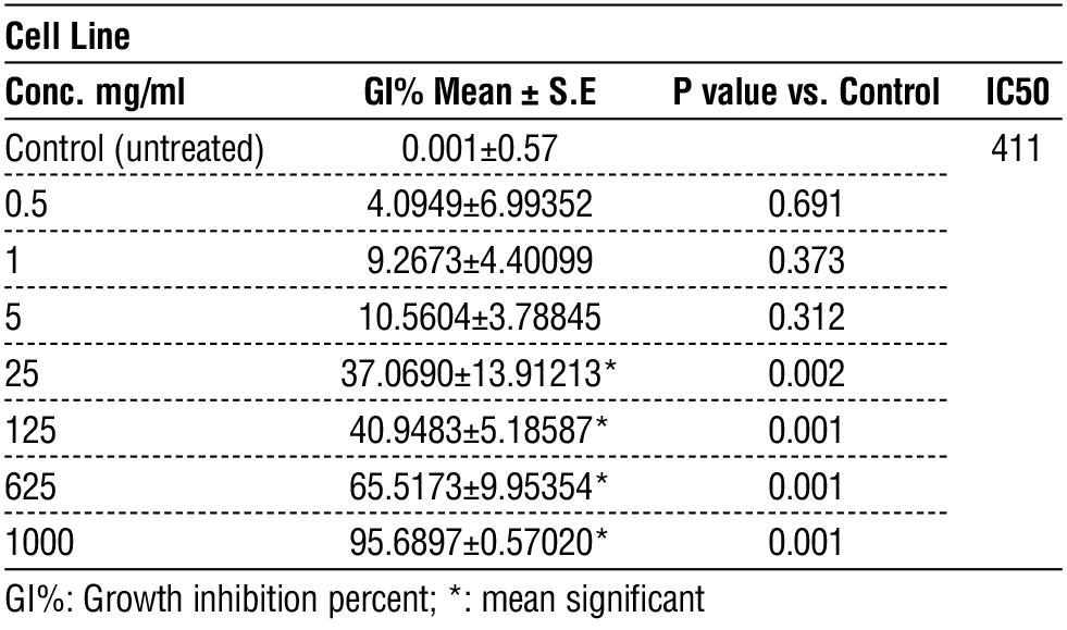
Table 1 - Cytotoxicity of MEX2R in LST174 cell line
 Results
Effect of MEX2R cytotoxicity on LST174 cell line
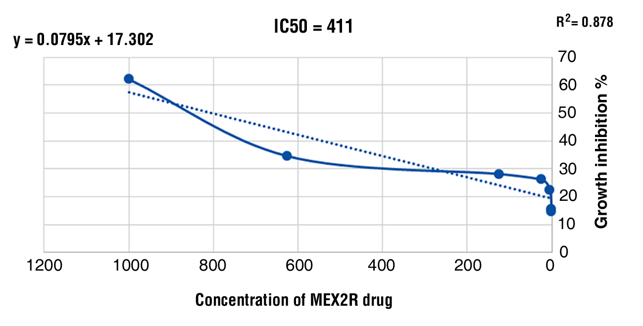
The results of the study showed that when comparing MEX2R at lower concentrations (0.5,1,5 ?g/ml) with the control, there was no significant variation (P > 0.05) in the inhibition of cell growth, while at other concentrations (25, 125,625 ?g/ml) of MEX2R, there was a highly significant increase (P ? 0.05) in the inhibition of LST174 cell growth when these concentrations were used for approximately 24 h of treatment and compared with the control group. In addition, the results showed that the concentration of drug required to inhibit a biological process or response by 50% (IC50) was approximately 411 mg/ml, as shown in table 1 and fig. 1.
Results
Effect of MEX2R cytotoxicity on LST174 cell line
The results of the study showed that when comparing MEX2R at lower concentrations (0.5,1,5 ?g/ml) with the control, there was no significant variation (P > 0.05) in the inhibition of cell growth, while at other concentrations (25, 125,625 ?g/ml) of MEX2R, there was a highly significant increase (P ? 0.05) in the inhibition of LST174 cell growth when these concentrations were used for approximately 24 h of treatment and compared with the control group. In addition, the results showed that the concentration of drug required to inhibit a biological process or response by 50% (IC50) was approximately 411 mg/ml, as shown in table 1 and fig. 1.
Figure 1 - Dose-response curve of growth inhibition of MEX2R on LST174 cell line presented by plotting drug concentrations versus growth inhibition %
Effect of MEX2O Cytotoxicity on LST174 Cell Line
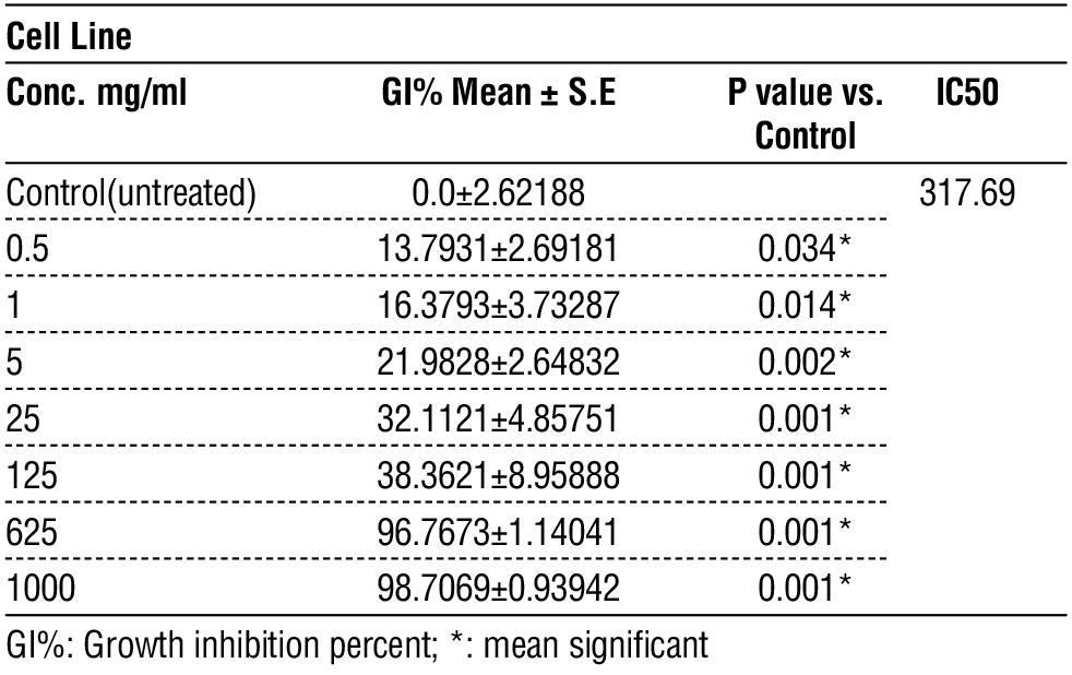
The result showed that MEX2O has significant difference (P?0.05) in LST174 cell growth inhibition at all concentrations (0.5,1.5.25,125,625 & 1000 ?g/ml) compared with the control(untreated) group. In addition, the results showed that the concentration of drug required to inhibit a biological process or response by 50% (IC50) was approximately 317.69 mg/ml as shown in table 2 and fig. 2.
Table 2 - Cytotoxicity of MEX2O in LST174 cell line
Effect of MTX Cytotoxicity on LST174 Cell Line
The results showed that MTX significantly increased (P ? 0.05) the growth inhibition of LST174 cells at all concentrations (0.5,1,5,25,125,625,1000 ?g/ml) compared with the control group. In addition, the results showed that the concentration of drug needed to inhibit a biological process or response by 50% (IC50) is approximately 803.8 mg/ml as in table 3 and fig. 3.
Figure 2 - Dose-response curve of growth inhibition of MEX2O on LST174 cell line presented by plotting drug concentrations versus growth inhibition %
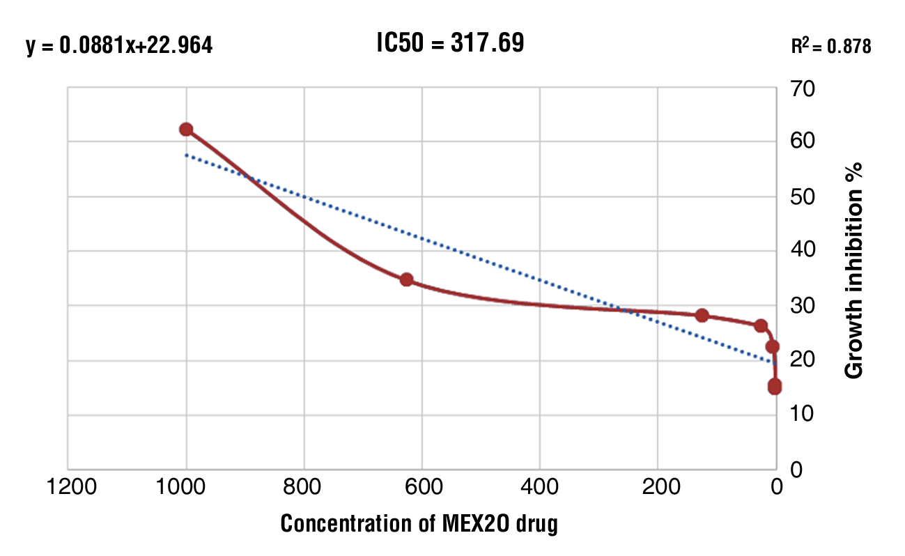
Table 3 - Cytotoxicity of MTX in LST174 cell line
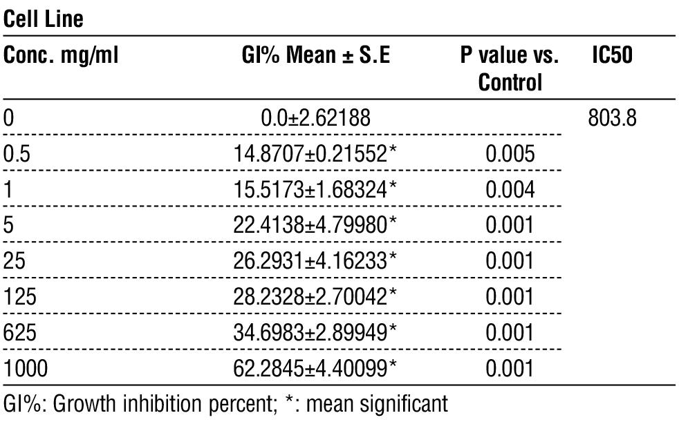
Figure 3 - Dose-response curve of growth inhibition of M on LST174 cell line presented
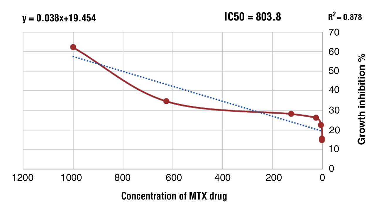
by plotting drug concentrations versus growth inhibition %
Discussion
KRAS is one of the most frequently mutated oncogenes in human cancer. To making this gene has a high priority therapeutic target so, most of KRAS mutant cancers are depended on the sustained expression and signaling of KRAS. However, the development of small molecules that directly inhibit KRAS function remains a challenge (18). There are many alternative therapeutic strategies used for KRAS mutant malignancies that involve targeting codependent vulnerabilities or synthetic lethal partners essential for oncogenic KRAS (19).
During the last two decades, many strategies have been developed to target oncogenic KRAS signaling. These strategies include the development of the KRAS protein direct inhibitors, usage of RNA interference strategies in addition to development of inhibitors act by prevent the localization of RAS to the plasma membrane, and pharmacologic targeting of its downstream effectors (20). Challenges and difficulties faced when designing a direct inhibitor of KRAS so, many earlier attempts to target KRAS-mutated CRC focused on inhibition upstream and downstream of KRAS signaling targets. Protein localization inhibitors, cyclin-dependent kinase (CDK) inhibitors, MEK inhibitors, focal adhesion kinase (FAK) inhibitors, and prenyl binding protein (PDE) inhibitors have been studied with varying degrees of success (21).
As previously discussed, one of the main mechanisms that is essential for localizing KRAS protein to the inner surface of the plasma membrane is farnesylation of KRAS, which transduces the signal from EGFR to RAF. Farnesyl transferase inhibitors (FTI) were developed as early KRAS blocking agents, and their function is to prevent the farnesylation of KRAS and block its migration to the cell membrane and sub sequent signaling (22). Salirasib is an oral RAS farnesyl cysteine mimetic that competitively blocks the membrane association of RAS proteins. In a phase II trial in advanced KRAS mutated NSCLC, it failed to provide any significant response and it cause many of adverse effects, the major adverse effects associated with the use of salirasib included diarrhea, nausea, and fatigue (23). Other agents such as Lonafarnib, which is currently approved to reduce the risk of death due to Hutchinson–Gilford progeria syndrome, and tipifarnib also failed to demonstrate any significant benefit in multiple phase II and phase III studies, which may be due to the presence of the bypass prenylation pathway via geranylgeranylation (24).
In this study, the results showed that MEX2R and MEX2O agents have highly significant cytotoxic effects on the LST174 colorectal cancer cell line when compared with MTX. These agents are effective in inhibiting biological processes or responses by 50% at doses less than the dose of MTX to produce the same effect, about half or less; IC50 for MEX2R & MEX2O (411 & 317.69 respectively). While the IC50 for MTX is approximately t 803.8, this means n that MTX is effective only at n high doses, e and thus a high concentration indicates n high systemic side effects. A previous study showed thate MTX caused e inhibition of o the dihydrofolate reductase enzyme and resulted t in the g inhibition of cell growth in SW480 cell, s which are e one of the colorectal cell lines that were tested in the study (25. However), t the anti proliferative effect of MTX may be deepened by n exposure time rather than the concentration of drug, s as found d by Huysentruy, et al., who s found that e MTX is less effective than cisplatin n on (VM M3) cultured cells and implanted mice (26).
Conclusion
From the results of this study, it can be concluded that MEX2R and MEX2O may have a more valuable role in this type of cancer treatment than MTX. These agents direct the cell cycle by targeting the KRAS gene mutation, which is one of the main obstacles that affects colon cancer treatment and hence inhibits cell proliferation.
Conflict of Interest
The authors have no conflict of interest.
Funding
There is no funding.
Ethical Consideration
This study was approved by the Ethical Committee of the Faculty, Medicine University of Kufa (June 2024, in accordance with document number 84).
References
1. Torre LA, Bray F, Siegel RL, Ferlay J, Lortet-Tieulent J, Jemal A. Global cancer statistics, 2012. CA Cancer J Clin. 2015;65(2):87-108.
2. Sajesh BV, Cisyk AL, McManus KJ. Synthetic Genetic Approaches in Colorectal Cancer: Exploiting and Targeting Genome Instability. 20th edn. Edited by C. Maxwell and C. Roskelley. Switzerland: Springer. 2015.
3. Victoir B, Croix C, Gouilleux F, Prié G. Targeted Therapeutic Strategies for the Treatment of Cancer. Cancers (Basel). 2024; 16(2):461.
4. Cox AD, Fesik SW, Kimmelman AC, Luo J, Der CJ. Drugging the undruggable RAS: Mission possible? Nat Rev Drug Discov. 2014; 13(11):828-51.
5. Guo F, Gong H, Zhao H, Chen J, Zhang Y, Zhang L, et al. Mutation status and prognostic values of KRAS, NRAS, BRAF and PIK3CA in 353 Chinese colorectal cancer patients. Sci Rep. 2018;8(1): 6076.
6. Xie MZ, Li JL, Cai ZM, Li KZ, Hu BL. Impact of primary colorectal Cancer location on the KRAS status and its prognostic value. BMC Gastroenterol. 2019;19(1):46.
7. Lim SM, Westover KD, Ficarro SB, Harrison RA, Choi HG, Pacold ME, et al. Therapeutic targeting of oncogenic K-Ras by a covalent catalytic site inhibitor. Angew Chem Int Ed Engl. 2014; 53(1):199-204.
8. Holstein SA, Hohl RJ. Is there a future for prenyltransferase inhibitors in cancer therapy? Curr Opin Pharmacol. 2012;12(6):704-9.
9. Asati V, Mahapatra DK, Bharti SK. K-Ras and its inhibitors towards personalized cancer treatment: Pharmacological and structural perspectives. Eur J Med Chem. 2017 Jan 5;125:299-314.
10. Scott AJ, Lieu CH, Messersmith WA. Therapeutic Approaches to RAS Mutation. Cancer J. 2016;22(3):165-74.
11. Ihle NT, Byers LA, Kim ES, Saintigny P, Lee JJ, Blumenschein GR, et al. Effect of KRAS oncogene substitutions on protein behavior: implications for signaling and clinical outcome. J Natl Cancer Inst. 2012;104(3):228-39.
12. Williams VL, Cohen PR, Stewart DJ. Sorafenib-induced premalignant and malignant skin lesions. Int J Dermatol. 2011;50(4):396-402.
13. Singh H, Longo DL, Chabner BA. Improving Prospects for Targeting RAS. J Clin Oncol. 2015;33(31):3650-9.
14. Bahrami A, Hassanian SM, ShahidSales S, Farjami Z, Hasanzadeh M, Anvari K, et al. Targeting RAS signaling pathway as a potential therapeutic target in the treatment of colorectal cancer. J Cell Physiol. 2018;233(3):2058-2066.
15. Mills CC, Kolb EA, Sampson VB. Development of Chemotherapy with Cell-Cycle Inhibitors for Adult and Pediatric Cancer Therapy. Cancer Res. 2018;78(2):320-325.
16. Chan ES, Cronstein BN. Mechanisms of action of methotrexate. Bull Hosp Jt Dis. 2013;71 Suppl 1:S5-8.
17. Davies JE. The pharmacological basis of therapeutics. Occup Environ Med. 2007;64(8):e2.
18. Hong DS, Fakih MG, Strickler JH, Desai J, Durm GA, Shapiro GI, et al. KRASG12C Inhibition with Sotorasib in Advanced Solid Tumors. N Engl J Med. 2020;383(13):1207-1217.
19. Aguirre AJ, Hahn WC. Synthetic Lethal Vulnerabilities in KRAS-Mutant Cancers. Cold Spring Harb Perspect Med. 2018;8(8): a031518.
20. Bhattacharya S, Socinski MA, Burns TF. KRAS mutant lung cancer: progress thus far on an elusive therapeutic target. Clin Transl Med. 2015;4(1):35.
21. Parikh K, Banna G, Liu SV, Friedlaender A, Desai A, Subbiah V, et al. Drugging KRAS: current perspectives and state-of-art review. J Hematol Oncol. 2022;15(1):152.
22. Appels NM, Beijnen JH, Schellens JH. Development of farnesyl transferase inhibitors: a review. Oncologist. 2005;10(8):565-78.
23. Riely GJ, Johnson ML, Medina C, Rizvi NA, Miller VA, Kris MG, et al. A phase II trial of Salirasib in patients with lung adenocarcinomas with KRAS mutations. J Thorac Oncol. 2011;6(8):1435-7.
24. Suzuki M, Jeng LJB, Chefo S, Wang Y, Price D, Li X, et al. FDA approval summary for lonafarnib (Zokinvy) for the treatment of Hutchinson-Gilford progeria syndrome and processing-deficient progeroid laminopathies. Genet Med. 2023;25(2):100335.
25. Morales C, Ribas M, Aiza G, Peinado MA. Genetic determinants of methotrexate responsiveness and resistance in colon cancer cells. Oncogene. 2005;24(45):6842-7.
26. Huysentruyt LC, Shelton LM, Seyfried TN. Influence of methotrexate and cisplatin on tumor progression and survival in the VM mouse model of systemic metastatic cancer. Int J Cancer. 2010;126(1):65-72.
Full Text Sources:

Abstract:

Views: 917


For Authors
Journal Subscriptions

Mar 2026
Supplements
Instructions for authors
Online submission
Contact
e-ISSN: 2601 - 1700 (online)
ISSN-L: 2559 - 723X
Journal Abbreviation: Surg. Gastroenterol. Oncol.
Surgery, Gastroenterology and Oncology (SGO) is indexed in:
- SCOPUS
- EBSCO
- DOI/Crossref
- Google Scholar
- SCImago
- Harvard Library
- Open Academic Journals Index (OAJI)
Surgery, Gastroenterology and Oncology (SGO) is an open-access, peer-reviewed online journal published by Celsius Publishing House. The journal allows readers to read, download, copy, distribute, print, search, or link to the full text of its articles.
Time to first editorial decision: 25 days
Rejection rate: 61%
CiteScore: 0.2
 IASGO Society News
Meetings and Courses in 2025
Meetings and Courses in 2024
Meetings and Courses in 2023
Meetings and Courses in 2022
Meetings and Courses in 2021
Meetings and Courses in 2020
Meetings and Courses in 2019
Verona expert meeting 2019

Surgery, Gastroenterology and Oncology applies the Creative Commons Attribution Non Commercial (CC BY-NC 4.0) license, which permits readers to copy and redistribute the material in any medium or format, remix, adapt, build upon the published works non-commercially, and license the derivative works on different terms, provided the original material is properly cited and the use is non-commercial. Please see: https://creativecommons.org/licenses/by-nc/4.0/
Publisher’s Note:
The opinions, statements, and data contained in article are solely those of the authors and not of Surgery, Gastroenterology and Oncology journal or the editors. Publisher and the editors disclaim responsibility for any damage resulting from any ideas, instructions, methods, or products referred to in the content.