Surgery, Gastroenterology and Oncology
|
|
Background: Cerebral edema is a critical complication following traumatic brain injury (TBI), often precipitating elevated intracranial pressure, cerebral ischemia, and subsequent cellular necrosis. Aquaporin-4 (AQP-4), an astrocytic water channel protein, plays a pivotal role in brain water homeostasis. Its upregulated expression following head trauma is frequently observed alongside water efflux into the brain interstitial tissue, a process closely linked to cerebral edema. This study aims to evaluate the association between serum AQP-4 levels and the severity of cerebral edema as assessed by head Computed Tomography (CT) scans in patients with TBI.
Methods: This was a quantitative analytical observational study with a cross-sectional design, conducted at Wahidin Sudirohusodo General Hospital from November to December 2023. A total of 70 TBI patients were included as respondents. Data on AQP-4 levels and CT scan-derived edema scores were collected and analyzed using the Spearman's rank correlation test.
Results: The study population was predominantly male (70%), with the most frequent age group being 12-25 years old (40%), and traffic accidents as the leading mechanism of injury (90%). Statistical analysis revealed a significant positive correlation between AQP-4 levels and the degree of cerebral edema on CT scans (p-value = 0.001), with a moderate correlation coefficient (r = 0.5).
Conclusion: Elevated serum AQP-4 levels are is significantly associated with the incidence and severity of cerebral edema in TBI patients. While these correlational clinical findings support the potential utility of circulating AQP-4 as a prognostic biomarker, further molecular studies are warranted to fully elucidate the underlying pathophysiological mechanisms and its exact relationship with local tissue expression.
Keywords: Aquaporin-4, cerebral edema, CT imaging, traumatic brain injury
INTRODUCTION
TBI is one of the leading causes of mortality and morbidity in the world (1). Globally, the incidence of brain injuries is increasing sharply mainly due to the increase in motor vehicle use dominated by adolescents and adults, making brain injuries the highest cause of death in productive ages 15-44 years (2). According to the Centers for Disease Control and Prevention (CDC) in 2019, approximately 166 people died from brain injuries in the United States every day (1).
Based on 2018 Basic Health Research, brain injuries ranked third in Indonesia, behind injuries to the lower and upper limbs, at 11.9% (2). Data from Wahidin Sudirohusodo General Hospital Makassar in 2021 show that there were 22 people (32.9 years old) between January 1 and December 31, 2021, followed by 11 people (16.5%), 10 people (16.5%), 10 people (14.9%), and 9 people (13.4%) who were between the ages of 31 and 40. There is one patient (1.5%), more males than females, in the age category of 61 to 65 years, which is the least represented in terms of patients (2).
Primary brain injury can progress to secondary brain injury characterized by cerebral edema. If cerebral edema in brain injury is not properly managed, it can lead ischemia in brain cells leading to death. Cerebral edema is the increase in brain volume due to elevated levels of water and sodium. Cerebral edema is the increase in brain volume due to elevated levels of water and sodium (3,4). AQP is a group of hydrophilic proteins known for their role in water transportation under both physiological and pathological conditions (5). AQP, particularly the AQP4 type, is predominantly expressed in astrocytes surrounding capillaries as well as in astrocyte processes consisting of glial limiting membranes and ependymal cells. It also plays a role in the processes and healing of cerebral edema. AQP is also involved in maintaining fluid balance in the central nervous system (CNS) and plays roles in signal transduction, neurogenesis, and neuroinflammation. In the context of trauma, elevated serum AQP-4 has emerged as a significant circulating biomarker associated with the clinical presentation of cerebral edema (6,7).
Edema is a ubiquitous response to various brain insults and may be cytotoxic, vasogenic, interstitial, or mixed in origin. Identifying the predominant imaging pattern, alongside clinical history, is crucial for accurate diagnosis and management. Computed tomography (CT) serves as the primary screening modality for patients presenting with acute neurological deficits. On a CT scan, edema manifests as a focal or diffuse area of hypoattenuation relative to normal brain parenchyma (8). Given this context, this study aims to investigate the correlation between serum AQP-4 levels and the radiographic severity of cerebral edema in TBI patients.
METHODS
This research is an observational quantitative analytic study with a cross-sectional research design. The research was conducted at Wahidin Sudirohusodo General Hospital, one of the largest central and eastern region hospitals in Indonesia, and also the main Teaching Hospital in South Sulawesi Province. As the primary tertiary trauma hub for this vast geographic region, our emergency department manages a continuously high daily volume of acute TBI admissions. The population in this study consists of all patients with brain injuries treated at Wahidin Sudirohusodo General Hospital. Due to this high influx of trauma cases, we were able to successfully recruit a sample size of 70 respondents using consecutive sampling over a concentrated two-month period from November to December 2023.
Inclusion Criteria
The inclusion criteria for the study are all patients with brain injuries treated at Wahidin Sudirohusodo General Hospital who are willing to participate in the study until completion.
Exclusion Criteria
The exclusion criteria for this study are blood serum samples that are damaged before AQP-4 examination, incomplete brain CT scan data, and patients lost during follow-up.
The variables in this study are AQP-4 levels as independent variables and the degree of edema assessed from the appearance of edema on brain CT scans as dependent variables. Blood samples for AQP-4 analysis and initial non-contrast head CT scans were routinely obtained upon patient admission to the emergency department. However, the exact time elapsed from the initial injury to clinical sampling was not strictly recorded or standardized due to variable pre-hospital transport times across our regional referral network. The degrees of cerebral edema are divided into 5 categories, namely: 0) No cerebral edema; 1) Focal edema limited to 1 lobe; 2) Unilateral edema (>1 lobe); 3) Bilateral edema; and 4) Global edema (disappearance of sulcal relief).
Statistical Analysis
The collected data were subjected to statistical analysis using the Spearman test.
RESULT
Characteristics of Study Respondents
The demographic and injury characteristics of the 70 TBI patients included in this study are summarized in table 1. The majority of respondents were male (n=49, 70.0%), reflecting the higher incidence of TBI in this sex. The most affected age group was teenagers (12-25 years), accounting for 40.0% (n=28) of the cohort. Consistent with global TBI epidemiology, motor vehicle crashes were the predominant mechanism of injury, responsible for 90.0% (n=63) of cases.
Table 1 - Characteristics of study respondents
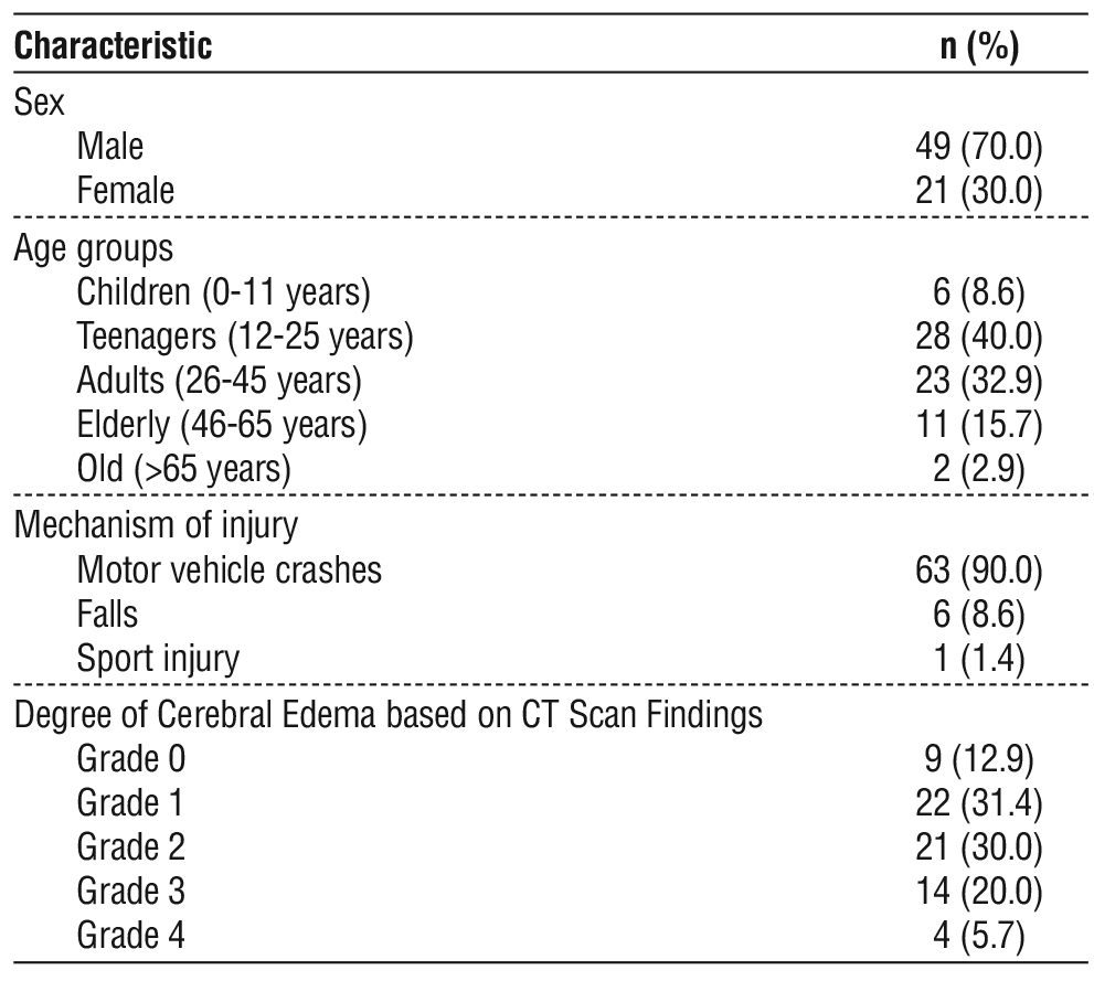
Regarding the severity of cerebral edema as assessed by CT scan, Grade 1 edema was the most prevalent (n=22, 31.4%), followed closely by Grade 2 (n=21, 30.0%). Notably, only a small minority of patients presented with Grade 4 global cerebral edema (n=4, 5.7%).
Characteristics of AQP-4 Levels
The quantitative characteristics of Aquaporin-4 (AQP-4) levels in the study population are presented in table 2. The mean AQP-4 level was found to be 1.818 ng/mL, with a standard deviation of 1.840 ng/mL. The 95% confidence interval for the mean AQP-4 level ranged from 1.38 to 2.26 ng/mL. Individual AQP-4 measurements spanned a considerable range, from a minimum of 0.184 ng/mL to a maximum of 6.901 ng/mL, indicating variability in AQP-4 expression among the TBI patients.
Table 2 - Characteristics of AQP-4 levels

Correlation Between AQP-4 Levels and Cerebral Edema Severity
The statistical analysis examining the association between AQP-4 levels and the degree of cerebral edema, as determined by CT scan findings, is displayed in table 3. A statistically significant positive correlation was identified between AQP-4 levels and the severity of cerebral edema (Spearman's ρ = 0.500; p < 0.001). This moderate positive correlation suggests that as AQP-4 levels increase, there is a tendency for the degree of cerebral edema on CT scans to also increase in TBI patients.
Table 3 - Correlation between AQP-4 levels and cerebral edema severity

DISCUSSION
Globally, traumatic brain injury becomes a significant health and socioeconomic issue. People of all ages are affected by it, and it is prevalent in both high and low-income nations (9). TBI is a serious and expensive global public health issue; an estimated 69 million new cases are reported each year, with a $400 billion USD global cost (10). The frequency of TBI is increasing, with a disproportionate rise in lower and middle-income nations (LMICs). By 2020, according to World Health Organization predictions, it will rank as the third leading cause of death and disability (11).
Based on sex characteristics, there were 49 (70%) male respondents and 21 (30 %) female respondents. Males were the majority of respondents in this study. The outcomes are consistent with the Kessely et al study showed the predominance was male (68.8%; n = 176) with a sex ratio of 2.2. 21.9% (n = 56) of patients were from rural areas (12). TBIs mostly occur in males because males are more physically active compared to females. Additionally, males also exhibit behaviors that tend to be more prone to injury (13).
The age group characteristics in this study show that adolescents (aged 12-25 years) are the largest group with a total of 28 respondents (40 %), followed by adults (aged 26-45 years) with 23 respondents (32.9 %), the elderly (aged 46-65 years) with 11 respondents (15.7 %), children (aged 6-12 years) with 6 respondents (8.6 %), and the least represented is the old (age above 65 years) with 2 respondents (2.9%). These results are consistent with the study by Danielle van Pelt et al., which found that the incidence of brain injuries in the population under 25 years old is highest in the age group of 12-18 years. The high incidence of brain injuries in the age range of 15-60 years is due to the busy lifestyle or high mobility level of individuals in that age group (14).
The primary mechanism causing brain injuries in this study is motor vehicle crashes (MHC), accounting for 63 cases (90%), followed by falls with 6 cases (8.6 %), and sports injuries with 1 case (1.4%). This investigation aligns with the work carried out by Hassan et al. that found that the commonest cause of TBI were MHC in 45% cases followed by falls 34% (15). In developed countries motor vehicle drivers are the most common victims whereas in developing countries, pedestrians and motorcyclist are the common victims (16). Motor vehicle crashes especially involving motorcyclist are the leading cause of morbidity and mortality (17). The safety surveillance programmes can certainly reduce the number of trauma patients as evident from the traffic safety regulations in Taiwan where implementation of the motorcycle helmet law decreased the incidence of motorcycle-related TBI (15).
In this study, the results indicate a significant relationship between the levels of AQP-4 and the level of edema based on CT scan findings (p-value < 0.001) and a moderate correlation (r-value = 0.500). It is important to note that while extensive experimental and animal models in the literature suggest a mechanistic and causal role for AQP-4 in driving fluid shifts and edema formation, our cross-sectional clinical findings specifically demonstrate a significant correlation rather than direct causality. Furthermore, a critical distinction must be made between local astrocytic AQP-4 expression and the circulating serum AQP-4 concentrations measured in this cohort. While experimental models utilize immunohistochemistry to demonstrate localized AQP-4 upregulation at the injured blood-brain barrier (BBB), our study captures systemic serum levels. We hypothesize that elevated serum AQP-4 in TBI patients reflects the release of this astrocytic protein into the systemic circulation secondary to post-traumatic BBB disruption, as supported by Mestre et al (18). Therefore, while serum AQP-4 serves as a valuable clinical correlate for edema severity, it does not necessarily provide a direct reflection of local tissue expression.
We must acknowledge that interpreting the biological role of AQP-4 solely as a driver of edema is a physiological oversimplification. As highlighted in foundational mechanistic literature, including studies by Mestre et al. (18) and Zhang et al. (19), AQP-4 exhibits a complex, dual role that is highly context-dependent, varying significantly with both the type of edema and the specific temporal phase of the injury.
In the early stages of TBI, which are heavily characterized by cytotoxic edema, upregulated AQP-4 acts detrimentally by facilitating the rapid influx of water into astrocytes, thereby exacerbating intracellular swelling (20). Conversely, during the vasogenic phase of edema - where water accumulates in the extra-cellular space secondary to blood-brain barrier breakdown—AQP-4 plays a crucial, neuroprotective role. In this context, functional AQP-4 is required to facilitate the clearance and efflux of excess interstitial fluid into the paravascular spaces and systemic circulation (18,21).
Because our clinical methodology utilized a standard non-contrast CT grading system that cannot reliably differentiate between cytotoxic and vasogenic edema on a microscopic level, our observed positive correlation between elevated serum AQP-4 and overall edema severity represents a net clinical snapshot rather than a specific pathway mechanism. This biological nuance is essential; it implies that therapeutic interventions targeting AQP-4 must be precisely timed. For instance, inhibiting AQP-4 may be acutely beneficial to prevent cytotoxic swelling, but the same inhibition could prove highly detrimental in later phases when AQP-4 is desperately needed for vasogenic fluid clearance.
Experimental and clinical evidence consistently demonstrates the temporal dynamics of AQP-4 following brain injury. Zhang et al (19). observed that local AQP-4 expression begins to increase within four hours post-trauma, peaking around eight to fifteen hours. Similarly, another study found that serum AQP-4 levels surge between 15 hours and eight days post-injury, a timeframe that corresponds temporally with severe BBB compromise and interstitial edema (18,21,22). Furthermore, AQP-4 gene suppression in animal models has been shown to reduce edema symptoms 24 hours after ischemic injury, under-scoring its pivotal role as a major determinant of overall brain fluid volume (21).
Non-contrast CT remains the gold standard for acute TBI triage, offering rapid, sensitive detection of neurosurgical emergencies such as hemorrhages, herniation, and significant edema. It surpasses MRI in the acute setting due to its speed, fracture sensitivity, and lack of contraindications for patients with penetrating or blast injuries (23). Because post-traumatic cerebral edema is a major driver of intracranial hypertension and subsequent mortality - affecting over 60% of patients with mass lesions - early radiographic detection is paramount (24). Although subtle density changes in white matter [e.g., a drop of >5 Hounsfield Units (HU)] denote early vasogenic progression, these changes are difficult to discern visually, emphasizing the need for supplementary biological markers like AQP-4 to help correlate and gauge edema severity (25,26).
Several limitations must be acknowledged when interpreting the findings of this study. First, there was a lack of strict temporal standardization in our study protocol. Experimental evidence clearly indicates that AQP-4 expression is highly time-dependent following traumatic brain injury, typically upregulating within hours and peaking between 8 to 24 hours post-injury before gradually declining (19,21,22). In our cohort, blood sampling and CT imaging were performed immediately upon emergency admission. However, the exact time from the traumatic event to sampling was not systematically recorded or standardized due to logistical constraints and widely variable transport delays to our center. Because we did not utilize standardized temporal sampling windows, the observed variability in serum AQP-4 levels may partially reflect these differences in post-injury timing rather than the severity of cerebral edema alone.
Second, we acknowledge that the CT grading system utilized in this study (Grades 0-4) is a relatively crude, categorical visual scale. While this scoring system is practical for rapid clinical triage in emergency settings, it lacks the precision of modern quantitative neuroimaging. The reliance on this visual scale over more rigorous metrics - such as quantitative HU analysis, volumetric edema measurements, midline shift quantification, and direct intracranial pressure correlation - limits the granularity of our edema severity assessments. Future prospective studies must incorporate strictly standardized sampling windows, serial AQP-4 measurements, and advanced quantitative neuroimaging to accurately uncouple the effects of time from the pathological progression of cerebral edema.
Third, a critical limitation of our statistical metho-dology is the reliance on univariate analysis. We did not perform a multivariate or ordinal regression model to adjust for significant potential clinical confounders, such as the initial Glasgow Coma Scale score, the presence and volume of intracranial hematomas, prior surgical decompression, the administration of osmotherapy (e.g., mannitol or hypertonic saline), and patient age. Given our relatively modest sample size (n=70), attempting a comprehensive multivariate model would risk statistical overfitting. Consequently, we cannot definitively conclude whether elevated AQP-4 is an independent biomarker of cerebral edema or if it merely correlates with overall traumatic injury severity.
Finally, while the correlation between serum AQP-4 and cerebral edema is statistically significant, the moderate coefficient (ρ = 0.500) yields a coefficient of determination (r2) of 0.25. This indicates that serum AQP-4 levels explain only 25% of the variance in edema severity, underscoring that AQP-4 cannot currently serve as a standalone clinical biomarker. Additionally, our study did not perform a Receiver Operating Characteristic (ROC) curve analysis to evaluate the specific predictive diagnostic performance or cutoff thresholds for AQP-4. Therefore, our clinical findings must be interpreted strictly as preliminary and hypo-thesis-generating. Future studies utilizing larger sample sizes are essential to conduct the robust multivariate analyses required to adjust for these critical covariates and isolate the independent pathophysiological role of AQP-4.
CONCLUSION
Elevated circulating serum levels of Aquaporin-4 are significantly associated with the incidence and radiographic severity of cerebral edema in patients with traumatic brain injury. Given the observational nature of this study, these preliminary, hypothesis-generating findings represent a strong clinical correlation rather than a direct causal relationship. Furthermore, while serum AQP-4 shows promise as a peripheral biomarker reflecting potential blood-brain barrier compromise, its current clinical utility as a standalone diagnostic tool remains limited. Further longitudinal, predictive, and molecular studies are required to fully elucidate the precise pathophysiological mechanisms governing AQP-4 dynamics in the acutely injured human brain.
Author’s Contributions
Conceptualization: WA, AAI, DW, NAL, WH, ARB, RG, and MF; Data curation: WA, AAI, DW, and NAL; Formal analysis: WA, AAI, and DW; Funding acquisition: WA, AAI, DW, NAL, and WH; Investigation: WA, AAI, DW, NAL, and WH; Methodology: WA, AAI, DW, NAL, and WH; Project administration: ARB, RG, and MF; Resources: ARB, RG, and MF; Software: ARB, RG, and MF; Supervision: WA, AAI, and DW; Validation: ARB, RG, and MF; Visualization: ARB, RG, and MF; Roles/Writing - original draft: WA, AAI, DW, NAL, WH, ARB, RG, and MF; and Writing - review & editing: WA, AAI, DW, and MF. All authors read and have approved the final version of the manuscript.
Competing Interest
The authors declare that they have no known competing financial interests or personal relationships that could have appeared to influence the work reported in this paper.
Funding
This research did not receive any specific grant from funding agencies in the public, commercial, or not-for-profit sectors.
Ethical Statement
The study was approved by the Ethics Committee of Hasanuddin University, Makassar, Indonesia (Protocol No. UH23070498).
REFERENCES
1. Albrecht JS, Hirshon JM, McCunn M, Bechtold KT, Rao V, Simoni-Wastila L, et al. Increased rates of mild traumatic brain injury among older adults in US Emergency Departments, 2009-2010. J Head Trauma Rehabil. 2016;31(5):E1-7.
2. Wardoyo MS, Widodo D, Ihwan A, Prihantono, Kusuma MI, Hendarto J, et al. The relationship between different dosages of mannitol 20% and osmolarity, blood sugar serum, and coagulation factors in moderate brain injury patients with increased intracranial pressure. Medicina Clínica Práctica 2021;4 (Suppl 1):100235.
3. Stokum JA, Gerzanich V, Simard JM. Molecular pathophysiology of cerebral edema. J Cereb Blood Flow Metab. 2016;36(3):513-38.
4. Prasetyo E. The primary, secondary, and tertiary brain injury. Crit Care Shock 2020;23:4-13.
5. Kurniawan VR, Islam AA, Adhimarta W, Zainuddin AA, Widodo D, Nasrullah, et al. The role of diphenhydramine HCl on tumor necrosis factor-a levels in wistar rats with traumatic brain injury: An in vivo study. Ann Med Surg (Lond). 2022;81:104399.
6. García Ríos CA, Leon-Rojas JE. Aquaporin-4 in Stroke and Brain Edema-Friend or Foe? Int J Mol Sci. 2025;26(17):8178.
7. Gontko-Romanowska K, Zaba Z, Panieński P, Steinborn B, Szemień M, Łukasik-Głębocka M, et al. The assessment of risk factors forfebrile seizures in children. Neurol Neurochir Pol. 2017;51(6):454-458.
8. Ho ML, Rojas R, Eisenberg RL. Cerebral edema. AJR Am J Roentgenol. 2012;199(3):W258-73.
9. Peeters W, van den Brande R, Polinder S, Brazinova A, Steyerberg EW, Lingsma HF, et al. Epidemiology of traumatic brain injury in Europe. Acta Neurochir (Wien). 2015;157(10):1683-96.
10. Dewan MC, Rattani A, Gupta S, Baticulon RE, Hung YC, Punchak M, et al. Estimating the global incidence of traumatic brain injury. J Neurosurg. 2018;130(4):1080-1097.
11. Brown JB, Kheng M, Carney NA, Rubiano AM, Puyana JC. Geographical disparity and traumatic brain injury in America: Rural areas suffer poorer outcomes. J Neurosci Rural Pract. 2019;10(1): 10-15.
12. Canton Kessely Y, Sobdjolbo O, Njesada N, Toudjingar FG, Ngaringuem O, Li-Iyane Ouambi O, et al. Pediatric traumatic brain injury in chad: about 256 cases. Egyptian Journal of Neurosurgery 2024;39:3.
13. Moppett IK. Traumatic brain injury: assessment, resuscitation and early management. Br J Anaesth. 2007;99(1):18-31.
14. Daniëlle Van Pelt E, De Kloet A, Hilberink SR, Lambregts SAM, Peeters E, Roebroeck ME, et al. The incidence of traumatic brain injury in young people in the catchment area of the University Hospital Rotterdam, the Netherlands. European Journal of Paediatric Neurology 2011;15:519–26.
15. Hassan N, Ali M, Haq NU, Azam F, Khan S, Khan Z, et al. Etiology, clinical presentation and outcome of traumatic brain injury patients presenting to a teaching hospital of Khyber Pakhtunkhwa. J Postgrad Med Inst 2017;31(4):365-70
16. Roozenbeek B, Maas AIR, Menon DK. Changing patterns in the epidemiology of traumatic brain injury. Nat Rev Neurol. 2013; 9(4):231-6.
17. Khan I, Khan A, Aziz F, Islam M, Shafqat S. Factors associated with helmet use among motorcycle users in Karachi, Pakistan. Acad Emerg Med. 2008;15(4):384-7.
18. Mestre H, Hablitz LM, Xavier AL, Feng W, Zou W, Pu T, et al. Aquaporin-4-dependent glymphatic solute transport in the rodent brain. Elife. 2018;7:e40070.
19. Zhang C, Chen J, Lu H. Expression of aquaporin-4 and pathological characteristics of brain injury in a rat model of traumatic brain injury. Mol Med Rep. 2015;12(5):7351-7.
20. Shi Z, Zhang W, Lu Y, Lu Y, Xu L, Fang Q, et al. Aquaporin 4-Mediated Glutamate-Induced Astrocyte Swelling Is Partially Mediated through Metabotropic Glutamate Receptor 5 Activation. Front Cell Neurosci. 2017;11:116.
21. Zhou Z, Zhan J, Cai Q, Xu F, Chai R, Lam K, et al. The Water Transport System in Astrocytes–Aquaporins. Cells. 2022;11(16): 2564.
22. Freire MAM, Rocha GS, Bittencourt LO, Falcao D, Lima RR, Cavalcanti JRLP. Cellular and Molecular Pathophysiology of Traumatic Brain Injury: What Have We Learned So Far? Biology (Basel). 2023;12(8):1139.
23. Ikeshima-Kataoka H. Neuroimmunological Implications of AQP4 in Astrocytes. Int J Mol Sci. 2016;17(8):1306.
24. Zhang M, Cui Z, Cui H, Cao Y, Wang Y, Zhong C. Astaxanthin alleviates cerebral edema by modulating NKCC1 and AQP4 expression after traumatic brain injury in mice. BMC Neurosci. 2016; 17(1):60.
25. Assentoft M, Larsen BR, MacAulay N. Regulation and Function of AQP4 in the Central Nervous System. Neurochem Res. 2015;40(12): 2615-27.
26. Schweitzer AD, Niogi SN, Whitlow CT, Tsiouris AJ. Traumatic brain injury: Imaging patterns and complications. Neurochem Res. 2015; 40(12):2615-27.
27. Asih MW, Martadiani ED, Laksminingsih NS, Sulaiman YW, Trisnawati IGA. Reliability of cerebral edema interpretation on head computed tomography scan in mild and moderate traumatic brain injury. Bali Medical Journal 2021;10(3):1067–9.
28. Wintermark M, Sanelli PC, Anzai Y, Tsiouris AJ, Whitlow CT, Druzgal TJ, et al. Imaging evidence and recommendations for traumatic brain injury: Advanced neuro- and neurovascular imaging techniques. AJNR Am J Neuroradiol. 2015;36(2):E1-E11.
29. Kim H, Kim G-dong, Yoon BC, Kim K, Kim BJ, Choi YH, et al. Quantitative analysis of computed tomography images and early detection of cerebral edema for pediatric traumatic brain injury patients: Retrospective study. BMC Med. 2014;12:186.
Full Text Sources:

Abstract:

Views: 27


For Authors
Journal Subscriptions

Jun 2026
Supplements
Instructions for authors
Online submission
Contact
e-ISSN: 2601 - 1700 (online)
ISSN-L: 2559 - 723X
Journal Abbreviation: Surg. Gastroenterol. Oncol.
Surgery, Gastroenterology and Oncology (SGO) is indexed in:
- SCOPUS
- EBSCO
- DOI/Crossref
- Google Scholar
- SCImago
- Harvard Library
- Open Academic Journals Index (OAJI)
Surgery, Gastroenterology and Oncology (SGO) is an open-access, peer-reviewed online journal published by Celsius Publishing House. The journal allows readers to read, download, copy, distribute, print, search, or link to the full text of its articles.
Time to first editorial decision: 25 days
Rejection rate: 61%
CiteScore: 0.2
 IASGO Society News

Meetings and Courses in 2025
Meetings and Courses in 2024
Meetings and Courses in 2023
Meetings and Courses in 2022
Meetings and Courses in 2021
Meetings and Courses in 2020
Meetings and Courses in 2019
Verona expert meeting 2019

Surgery, Gastroenterology and Oncology applies the Creative Commons Attribution Non Commercial (CC BY-NC 4.0) license, which permits readers to copy and redistribute the material in any medium or format, remix, adapt, build upon the published works non-commercially, and license the derivative works on different terms, provided the original material is properly cited and the use is non-commercial. Please see: https://creativecommons.org/licenses/by-nc/4.0/
Publisher’s Note:
The opinions, statements, and data contained in article are solely those of the authors and not of Surgery, Gastroenterology and Oncology journal or the editors. Publisher and the editors disclaim responsibility for any damage resulting from any ideas, instructions, methods, or products referred to in the content.