Surgery, Gastroenterology and Oncology

|
|
Introduction: High-grade renal trauma has been previously associated with nephrectomy. Nevertheless, advancements in medicine have shifted management towards organ-preserving strategies in haemodynamically stable patients. Computed tomography (CT) plays a pivotal role in the initial diagnosis, trauma grading, therapy guiding and follow-up. The conservative approach is particularly beneficial in sports-related injuries, where patients are young and renal preservation provides significant long-term benefits.
Clinical Case: This article reviews the case of a previously healthy 18-year-old male who presented to the emergency department following a blunt renal trauma during a football game. He complained of right flank pain associated with macroscopic haematuria. On admission, he was haemodynamically stable. CT showed severe parenchymal disruption of the right kidney with urinoma and retroperitoneal haematoma. The findings correspond to Grade IV renal injury according to the American Association for the Surgery of Trauma (AAST). Considering the absence of haemodynamic compromise, the patient was managed conservatively with clinical follow-up.
Conclusion: This case aims to emphasise that a CT-guided conservative management in patients with grade IV renal trauma should be the preferred strategy in haemodynamically stable cases. Surgical intervention should be avoided whenever feasible, particularly in sports-related trauma, where patients are often young and organ-preserving strategies are of paramount importance.
Keywords: renal trauma, grade IV, CT, sports trauma, conservative management, urinoma, haemodynamic stability
INTRODUCTION
Renal trauma in sports is uncommon and accounts for 10-20% of all renal injuries. It typically results from direct blunt force to the flank. The most common causes are road traffic accidents and falls from height (1,2). The vast majority of these injuries are minor, although severe renal trauma can occur. While low-grade injuries are commonly treated conservatively, high-grade renal trauma remains clinically challenging due to possible complications such as haemorrhage, urinary extravasation and long-term renal impairment. Grade IV renal trauma is defined by AAST as 'a parenchymal laceration extending through the cortex and medulla into the urinary collecting system, causing urine leakage, or a vascular injury causing segmental infarction or active bleeding' (3). Its management historically favoured prompt operative intervention with high nephrectomy rates. However, the role of surgery in blunt solid abdominal organ trauma has diminished significantly with the evolution of critical care, minimally invasive endovascular techniques and especially high-resolution CT, which is not only used in diagnosing, but also plays an important role in treatment planning and follow-up (4).
Case Report
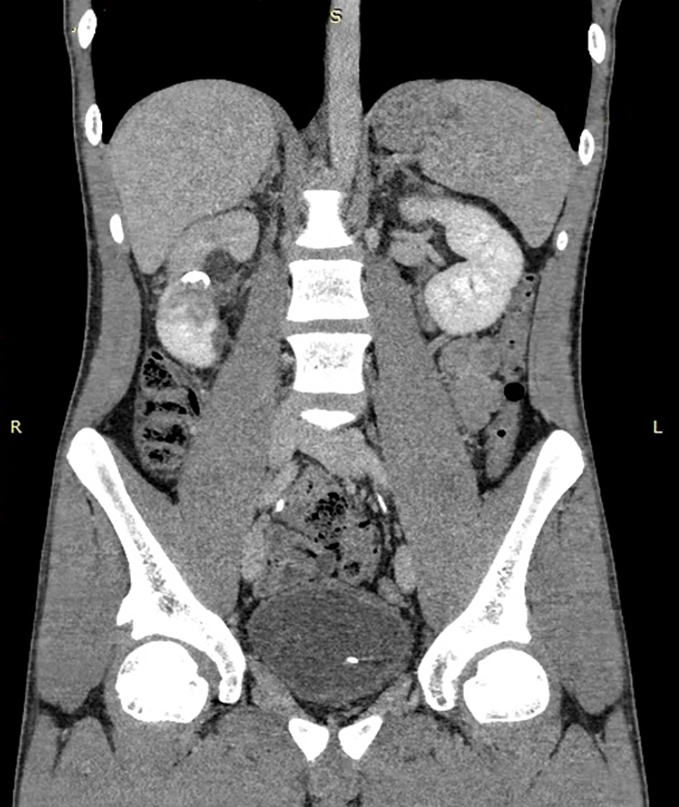
An 18-year-old male was brought to the emergency department with right flank pain and macroscopic haematuria following a ball strike during an amateur football game. He had no previous medical records. He was not taking any medications and had no known allergies. On examination, he was alert and oriented. His blood pressure and heart rate were within the normal range. His abdomen was soft, with no guarding or pain during palpation. Bowel sounds were present. Succusio renalis was bilaterally negative. Laboratory tests on admission showed haematuria and mild anaemia, with a haemoglobin level of 124 g/L that dropped to 102 g/L on the following day. eGFR remained relatively stable throughout the admission at around 70 mL/min. CT scan of the abdomen and pelvis, performed as a four-phase study with contrast, including delayed phase, showed deep right kidney laceration in the middle third with involvement of the collecting system and mixed urinary and blood extravasation in the retroperitoneal space - findings consistent with Grade IV renal injury (figs. 1, 2). In addition, suspicious findings included a small thrombus in the right renal vein. No other injuries were identified. Given the patient's haemodynamic stability, a conservative management strategy was adopted. He was placed on bed rest, and intravenous fluids were administered to maintain adequate perfusion. Antibiotics were given to cover for possible infection. He remained closely monitored for signs of worsening anaemia or haemodynamic deterioration during his admission. A JJ ureteral stent was placed via cystoscopy to facilitate urinary drainage. Follow-up CT performed 24h later showed persistent laceration of the right kidney with dilated upper group calyces and complete resorption of the previous fluid collections. The patient remained haemodynamically stable, without any new complaints, and was discharged on day 12 of admission. Four months later, he came for a follow-up, and he was found to be asymptomatic, with normal renal function. CT showed almost complete recovery of the kidney with dilated upper calyces and no perifocal fluid collections (fig. 3), and the ureteral stent was successfully removed.
Figure 1 - Contrast-enhanced CT at arterial phase in sagittal plane showing deep laceration of the parenchyma and extensive retroperitoneal heterogenous fluid collection
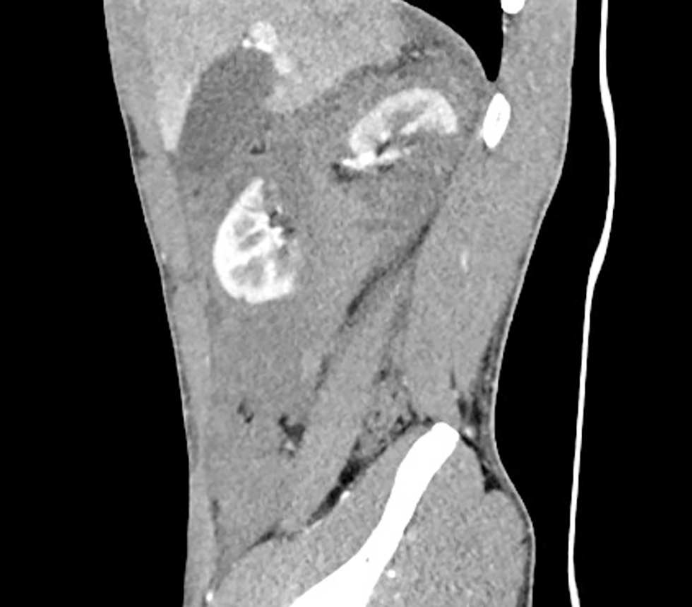
Figure 2 - Contrast-enhanced CT at urographic phase showing right kidney laceration and large amount of contrast extravasation in the retroperitoneal space corresponding to urinoma
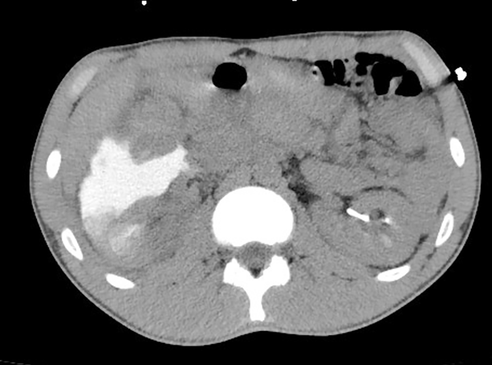
Figure 3 - Follow-up CT urography performed four months after the initial scan. Coronal urographic-phase image demonstrates a right-sided JJ ureteric stent with the proximal pigtail positioned within the right renal pelvis. Hypoenhancement of the upper pole renal parenchyma is noted, associated with dilatation of the upper calyceal system. The previously identified retroperitoneal fluid collections have completely resolved.
DISCUSSIONS
Football-related renal trauma is rare and could present a diagnostic challenge. The majority of kidney injuries from football have been noted in patients in the 16–18-year age group(5), which suggests that these patients are physically active and without significant comorbidities. This makes organ preservation particularly important. Moreover, the mechanism of injury is usually isolated blunt trauma, increasing the likelihood of successful conservative management (6). However, severe injuries still require careful multidisciplinary assessment and precise clinical observation due to the risk of delayed bleeding, urinoma formation, infection, or haemodynamic deterioration. Therefore, repeated clinical evaluation, laboratory monitoring, and follow-up imaging remain essential components of safe NOM.
This case emphasises the importance of contrast-enhanced CT in the diagnosis and management of high-grade renal trauma. CT allowed accurate grading of the injury and identification of complications such as urinary extravasation and haematoma. It also allows exclusion of active major vascular bleeding or associated intra-abdominal injuries requiring prompt surgical intervention. Exceeding its diagnostic value, CT also serves as a tool for serial assessment, providing close radiological monitoring of injury evolution and contributing to the conservative treatment strategy. In modern trauma care, CT has become the gold standard for initial injury characterisation, risk stratification, and long-term follow-up in haemodynamically stable patients (7).
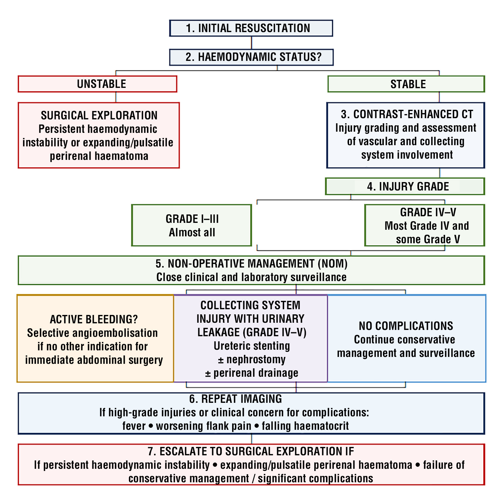
According to the EAU Guidelines (8), NOM is the preferred approach for most blunt renal injuries in stable patients, including selected Grade IV injuries. Patient selection is central to the success of conservative management in high-grade renal trauma. Current EAU recommendations identify haemodynamic stability as the primary criterion for a non-operative strategy. In addition to stable vital signs and an adequate response to initial resuscitation, the decision should incorporate the mechanism and grade of injury, transfusion requirements, associated intra-abdominal injuries, and CT findings. Radiological features associated with a higher likelihood of intervention include active vascular contrast extravasation, a large or expanding perirenal haematoma, and vascular injuries such as a pseudoaneurysm or arteriovenous fistula. On the other hand, a haemodynamically stable patient without ongoing major haemorrhage or another abdominal injury requiring surgical exploration may be considered for NOM even in the presence of a high-grade renal injury. This can avoid the morbidity associated with surgery, which is articularly important in young athletes, where renal preservation can avoid potential long-term consequences of nephrectomy, including an increased risk of hypertension and chronic kidney disease later in life (9). Nevertheless, Grade IV injuries require close clinical and laboratory monitoring because the likelihood of subsequent intervention is higher than in low-grade trauma.
Based on the AAST classification and current EAU recommendations, table 1 presents a simplified therapeutic algorithm for renal trauma. Haemodynamic stability remains the principal determinant of NOM, while angioembolisation, urinary drainage, or surgical exploration may be required based on clinical course and imaging findings.
Table 1 - Simplified algorithm for management of renal trauma
In our patient, the presence of collecting-system injury with urinary extravasation prompted JJ stent placement after the initial CT to facilitate internal urinary drainage. Follow-up CT demonstrated resolution of the retroperitoneal fluid collections, supporting continued conservative management.
The present case contributes to the growing evidence supporting CT-guided conservative management as a preferred strategy in selected patients with Grade IV sports-related renal trauma. Successful renal preservation is feasible even in severe trauma previously managed operatively, with contemporary strategies such as early imaging, a multidisciplinary approach, and careful monitoring (10).
Several limitations should be acknowledged. As a single case, this report cannot establish the superiority of NOM for all Grade IV renal injuries. In addition, the short follow-up in this case is insufficient to exclude delayed complications, including bleeding, urinary obstruction or infection, vascular complications, and post-traumatic hypertension. Therefore, long-term surveillance remains important after high-grade renal trauma.
CONCLUSIONS
This case demonstrates that NOM could be a safe alternative to surgery in carefully selected patients with Grade IV renal trauma who remain haemodynamically stable. Conservative management could be especially beneficial in young patients with sports-related trauma. Contrast-enhanced CT plays an essential role in accurate diagnosis and injury grading. Moreover, it guides the therapeutic management and the continuous assessment. Continued research is needed to further refine patient selection criteria for non-operative management of high-grade renal trauma.
Authorship
All authors confirm that they have met the criteria for authorship as established by the International Committee of Medical Journal Editors.
Conflicts of Interest and Source of Funding
The authors state no conflicts of interest and declare no funding was received for this study.
REFERENCES
1. Shin CI, Sim JS. Renal Trauma. In: KIM SH. (eds) Uroradiology. Singapore: Springer; 2025.
2. Saidi A, Bocqueraz F, Descotes JL, Cadi P, Terrier N, Boillot B, et al. Blunt kidney trauma: a ten-year experience. Prog Urol. 2004; 14(6):1125-31. French
3. Kozar RA, Crandall M, Shanmuganathan K, Zarzaur BL, Coburn M, Cribari C, et al. Organ injury scaling 2018 update: spleen, liver, and kidney. J Trauma Acute Care Surg. 2018;85(6):1119-1122.
4. Kautza B, Zuckerbraun B, Peitzman AB. Management of blunt renal injury: what is new? Eur J Trauma Emerg Surg. 2015;41(3): 251-8.
5. Kim JK, Koyle MA, Lee MJ, Nason GJ, Ren LY, O'Kelly F. A systematic review of genitourinary injuries arising from rugby and football. J Pediatr Urol. 2020;16(2):130-148.
6. Patel DP, Redshaw JD, Breyer BN, Smith TG, Erickson BA, Majercik SD, et al. High-grade renal injuries are often isolated in sports-related trauma. Injury. 2015;46(7):1245-9.
7. Dayal M, Gamanagatti S, Kumar A. Imaging in renal trauma. World J Radiol. 2013;5(8):275-84.
8. EAU Guidelines. Edn. presented at the EAU Annual Congress, London 2026.
9. American Urological Association. Urotrauma. 2017.
10. Bedoui MA, Saïdani B, Saadi A, Taher SB, Chakroun M, Slama RB. Conservative management of blunt high-grade renal trauma: A case report. Int J Surg Case Rep. 2025;128:111059.
Full Text Sources:

Abstract:

Views: 289


For Authors
Journal Subscriptions

Jun 2026
Supplements
Instructions for authors
Online submission
Contact
e-ISSN: 2601 - 1700 (online)
ISSN-L: 2559 - 723X
Journal Abbreviation: Surg. Gastroenterol. Oncol.
Surgery, Gastroenterology and Oncology (SGO) is indexed in:
- SCOPUS
- EBSCO
- DOI/Crossref
- Google Scholar
- SCImago
- Harvard Library
- Open Academic Journals Index (OAJI)
Surgery, Gastroenterology and Oncology (SGO) is an open-access, peer-reviewed online journal published by Celsius Publishing House. The journal allows readers to read, download, copy, distribute, print, search, or link to the full text of its articles.
Time to first editorial decision: 25 days
Rejection rate: 61%
CiteScore: 0.2
 IASGO Society News
Meetings and Courses in 2025
Meetings and Courses in 2024
Meetings and Courses in 2023
Meetings and Courses in 2022
Meetings and Courses in 2021
Meetings and Courses in 2020
Meetings and Courses in 2019
Verona expert meeting 2019

Surgery, Gastroenterology and Oncology applies the Creative Commons Attribution Non Commercial (CC BY-NC 4.0) license, which permits readers to copy and redistribute the material in any medium or format, remix, adapt, build upon the published works non-commercially, and license the derivative works on different terms, provided the original material is properly cited and the use is non-commercial. Please see: https://creativecommons.org/licenses/by-nc/4.0/
Publisher’s Note:
The opinions, statements, and data contained in article are solely those of the authors and not of Surgery, Gastroenterology and Oncology journal or the editors. Publisher and the editors disclaim responsibility for any damage resulting from any ideas, instructions, methods, or products referred to in the content.