Surgery, Gastroenterology and Oncology

|
|
Background: Considering the increasing incidence and mortality of colorectal cancer, MLH1 plays a critical role as an essential component of the DNA mismatch repair (MMR) system, ensuring genomic stability during cell division. Impairment of MLH1 function contributes to an increased susceptibility to colorectal cancer development. Objective: This study aimed to investigate the relationship between protein expression status and the demographic and histopathological characteristics of patients with colorectal cancer (CRC) and evaluate its impact on overall survival.
Methodology: Tissue samples preserved in paraffin were obtained from 100 patients
diagnosed with colorectal cancer in the Thi-Qar Governorate, Iraq. Corresponding clinical and pathological data, including demographic and histopathological features, were collected to assess their relationship with MLH1 protein expression.
Results: MLH1 protein expression was lost in 14% of the patients (14/100). Loss of
expression was significantly associated with age < 50 years (P = 0.007), whereas no
significant correlation was observed with sex or family history. Furthermore, MLH1 loss was significantly associated with the tumor histological type (P = 0.02), tumor location (P = 0.005), and T-stage (P = 0.006). Survival analysis using Kaplan–Meier curves and log-rank test revealed no significant difference in overall survival between patients with intact MLH1 expression and those exhibiting loss of expression (P = 0.197).
Conclusion: The results demonstrated an increased rate of MLH1 loss of expression in patients younger than 50 years, across different tumor types, as well as in right-sided colon tumors and advanced stages of colorectal cancer, with no statistically significant association observed with overall survival.
Keywords: colorectal cancer, immunohistochemistry, MLH1
Introduction
Colorectal cancer is one of the most prevalent malignancies worldwide and the third leading cause of cancer-related mortality globally. It is one of the most common malignant tumors affecting the digestive system (1). Colorectal cancer is a malignant neoplasm arising from the epithelial cells lining the colon and rectum through their transformation from normal to cancerous glandular cells (2). It is generally estimated that approximately 90% of colorectal cancers are acquired factors and are often related to lifestyle and dietary habits, while approximately 10% of these causes are due to inherited genetic predisposition (3). Three main pathways can arise and progress in cancer: chromosomal instability (CIN), microsatellite instability (MSI), and serrated pathways (4). The CIN pathway is characterized by mutations in tumor suppressor genes and oncogenes that drive adenoma – carcinoma sequences, such as APC, KRAS, and TP53. APC are among the most frequent mutations in approximately 80% of sporadic colorectal cancer cases (5). This pathway (MSI) represents 15% of colorectal cancer pathways and is characterized by recurrent mutations in new DNA base pairs, resulting in a hypermutator phenotype. This, in turn, is attributed to genetic and hereditary changes in MMR systems (6). One of the main causes of colorectal cancer is problems in the functioning of the mismatch repair system. Based on the relationship between the problems that occur in MMR and the causes that lead to them, colorectal cancer has been classified into different groups. The first group refers to sporadic colorectal cancer that suffers from a deficiency in mismatch repair caused by high methylation of the promoter region of the MLH1 gene, which leads to functional silencing of this gene in tumors that are affected by the methylation pattern of CPG islets (7). The second group of inherited colorectal cancers
suffer from a deficiency in mismatch repair genes, which results mainly from Lynch syndrome and is characterized by germline mutations in one or more of the basic mismatch repair system genes MLH1, PMS2, MSH2, or MSH6 (8,9).
Material and method
Sample Collection
The current study included (100) samples of formalin-fixed and paraffin-embedded colon and rectal cancer tissue samples. Samples were collected from patients undergoing surgical intervention at Imam Hussein Teaching Hospital in Thi-Qar Governorate, Iraq, between 2023 and 2025. Demographic data (age, sex, and family history) were obtained from the patient records at the Oncology Center of the Thi-Qar Governorate Health Department, Iraq. Histological variables (tumor type, tumor location, tumor grade, tumor stage, and lymph node metastases) were obtained by re-evaluation of the histological sections using routine staining by a histopathologist. The collection of samples and data related to the research was approved by the Scientific Research Ethics Committee of the Thi-Qar Health Department/Ministry of Health/Iraq (Approval No. 34/2025, Approval Date 1/20/2025).
Immunohistochemically Staining
Formalin-fixed, paraffin-embedded tissue sections were cut at 4 µm thickness and mounted on positively charged glass slides, deparaffinized in xylene, and rehydrated through graded alcohol solutions. Antigen retrieval was performed using EnVision™ FLEX Target Retrieval Solution,(Tris/EDTA buffer, pH 9; Dako, Denmark) in a conventional oven. Endogenous peroxidase activity was blocked using EnVision™ FLEX Peroxidase Blocking Reagent. The sections were then incubated with a ready-to-use mouse monoclonal anti-MLH1 antibody (Clone IR079,Dako,Denmark) according to the manufacturer's instructions. Immuno-reactivity was detected using the EnVision™ FLEX HRP/DAB Detection System (Dako, Denmark) and visualized with 3,3′-diaminobenzidine (DAB) chromogen. The slides were counterstained with hematoxylin,dehydrated,mounted,and examined using a Leica ICC50 light microscope.
MLH1 immunoreactivity was evaluated by an experienced pathologist using a semi-quantitative scoring system based on staining intensity and the proportion of positive tumor cells; only nuclear staining was considered positive. The staining intensity was scored as 0 (none), 1 (weak), 2 (moderate), and 3 (strong). The proportion of positive tumor cells was scored as 0 (none), 1 (<1%), 2 (1–10%), 3 (10–33%), 4 (33–66%), and 5 (66–100%). The final immunoreactivity score was calculated as the sum of the intensity and proportion scores. Cases with scores of 0–2 were classified as negative, whereas those with scores of 3–8 were classified as positive.
Statistical Analysis
An independent-samples t-test was used to compare continuous variables between study groups, with statistical significance set at P ≤ 0.05. All statistical analyses were performed using the SPSS software. Survival analysis was performed using the Kaplan-Meier method to estimate disease-free survival (DFS). Differences in survival between patients with intact MLH1 expression and those with loss of expression were assessed using the log-rank test.
Results
Clinical and Pathological Characteristics
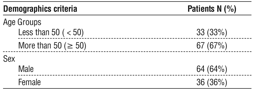
The results showed that The mean age of the patients was (55.70 ± 14.20) years. The patients were categorized into two age groups: those aged 50 years or older (n = 67, 67%) and those younger than 50 years (n = 33, 33%). Regarding sex distribution, males comprised 64% of the cohort, while females accounted for 36%, as summarized in table 1.
Table 1 - The clinical and pathological characteristics of colorectal cancer patients.
Determining the Expression Status of MLH1 Protein in Colorectal Cancer Patients
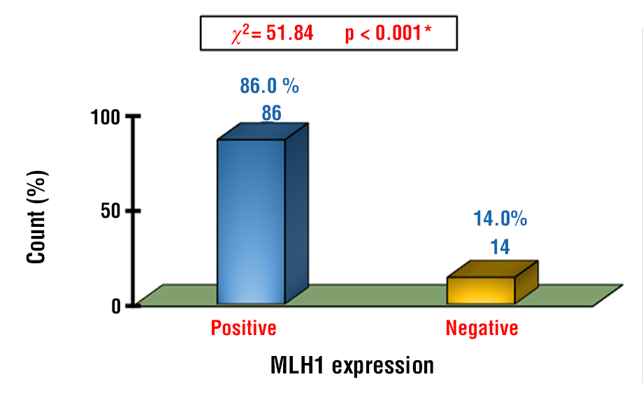
MLH1 protein expression was positive in 86% of the cases, whereas 14% of the cases exhibited loss of expression, with a statistically significant difference between the two groups, as illustrated in fig. 1.
Figure 1 - The expression status of MLH1 in CRC patients with colorectal cancer.
The Relationship Between MLH1 Protein Expression Status and Demographic Characteristics
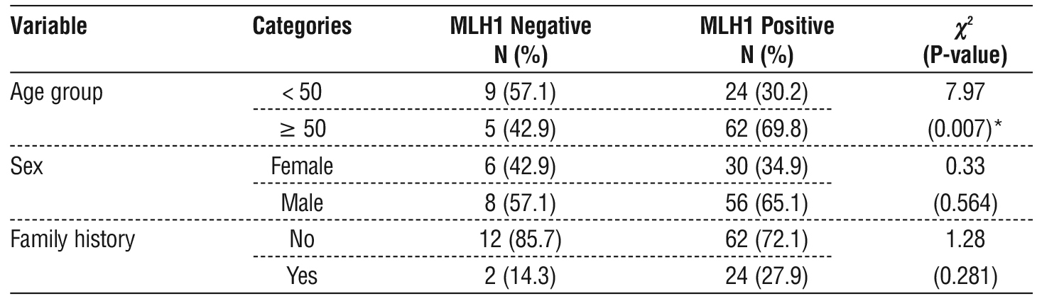
Loss of MLH1 protein expression was more frequent in patients aged < 50 years than in those aged ≥ 50 years, with a statistically significant association between expression status and age group. The frequency of MLH1 loss was also higher in males than in females; however, this difference was not statistically significant, and no significant association was observed between MLH1 expression status and family history, as most cases of MLH1 loss occurred in patients without a positive family history (table 2).
Table 2 - Association between MLH1 protein expression status and demographic variables in patients with colorectal cancer.
The Relationship Between MLH1 Protein Expression Status and Histological Variables
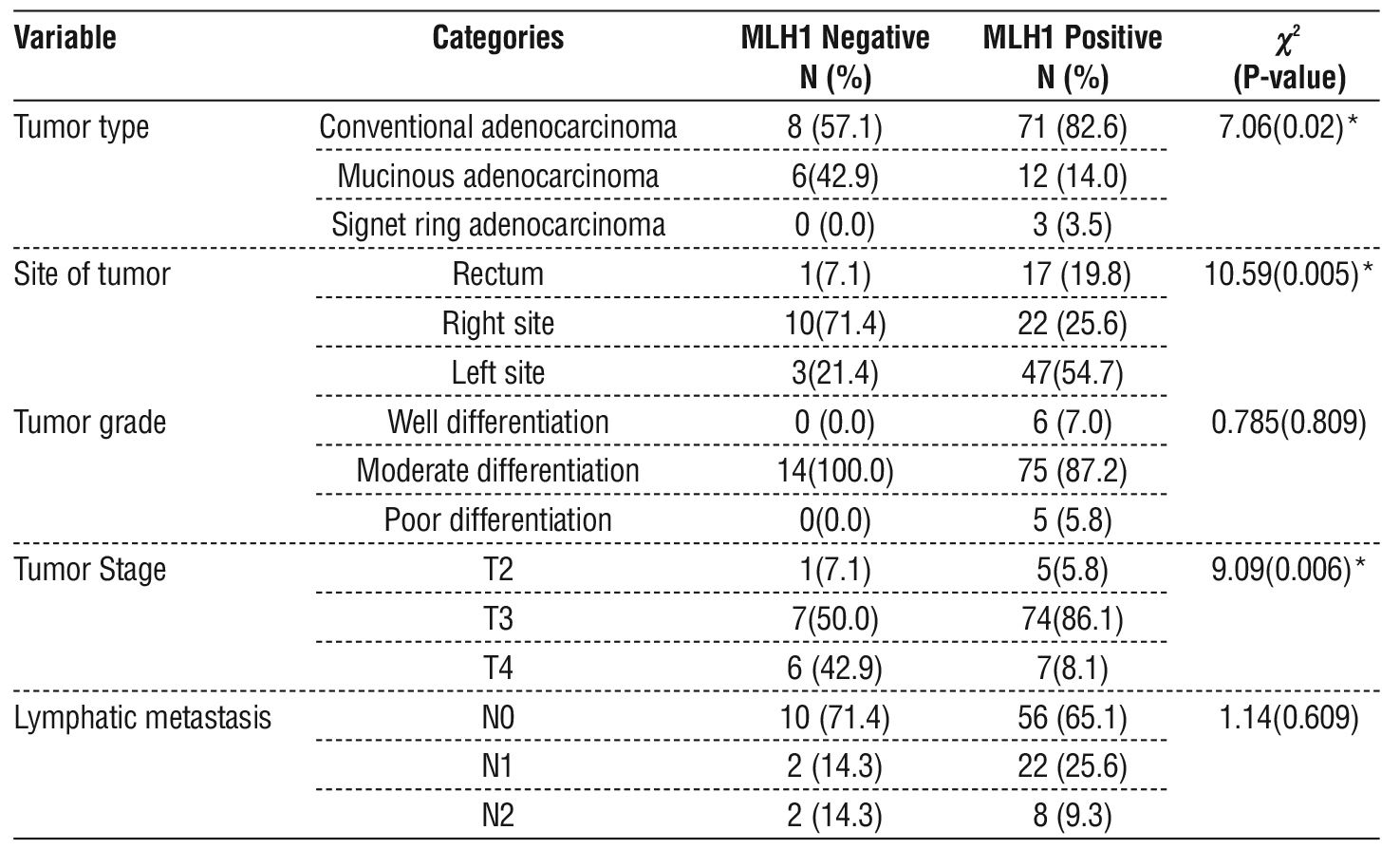
A significant association was observed between MLH1 protein expression status and histopathological tumor type. Loss of MLH1 expression was most frequently observed in conventional adenocarcinomas, followed by mucinous adenocarcinomas, whereas no loss of expression was detected in signet-ring cell carcinomas.
A significant association was observed between the MLH1 protein expression status and tumor location. Loss of MLH1 expression was most frequently detected in right-sided colon tumors, followed by left-sided colon tumors, whereas rectal tumors exhibited the lowest frequency of MLH1 loss. In contrast, no significant association was found between MLH1 expression status and tumor differentiation, although all cases of MLH1 loss were identified in moderately differentiated tumors. Furthermore, MLH1 protein expression status was significantly associated with tumor stage, with loss of expression occurring predominantly in advanced-stage tumors, particularly in T3 and T4 lesions.
No significant association was found between the MLH1 protein expression status and lymph node metastasis. Although the loss of MLH1 expression was more frequently observed in tumors without lymph node involvement, the distribution of MLH1 expression status across lymph node categories did not reach statistical significance (table 3).
Table 3 - The relationship between the expression status of MLH1 and histological variables in patients with colon and rectal cancer.
Effect of MLH1 Protein Expression Status on Disease-Free Survival
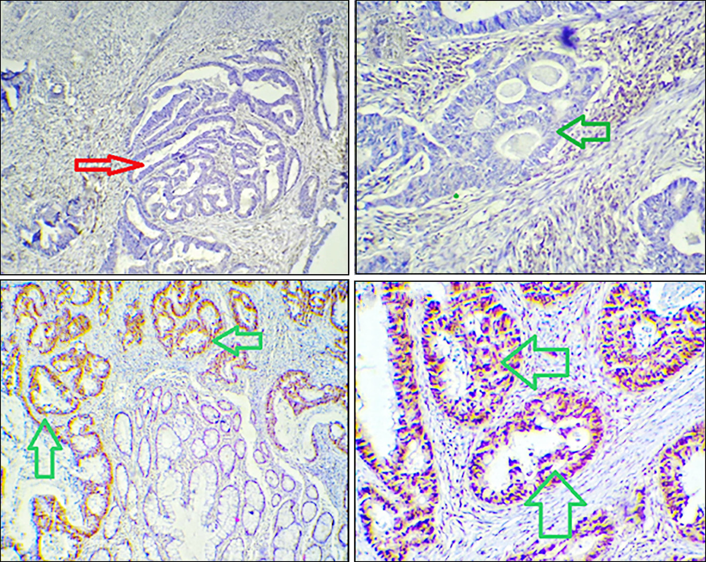
Patients with positive MLH1 protein expression exhibited longer overall survival than those with loss of expression. However, this difference was not statistically significant according to the log-rank test. Accordingly, no significant association was observed between MLH1 expression and overall survival in the present study (table 4, fig. 2). The histopathology and IHC results are shown in fig. 3.
Table 4 - The relationship between disease-free survival and MLH1 protein expression status.
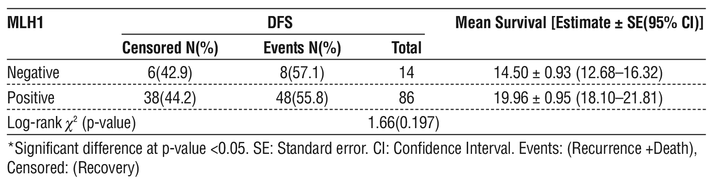
Figure 2 - The Kaplan-Meier curves for disease-free survival according to the expression status of the MLH1 protein.
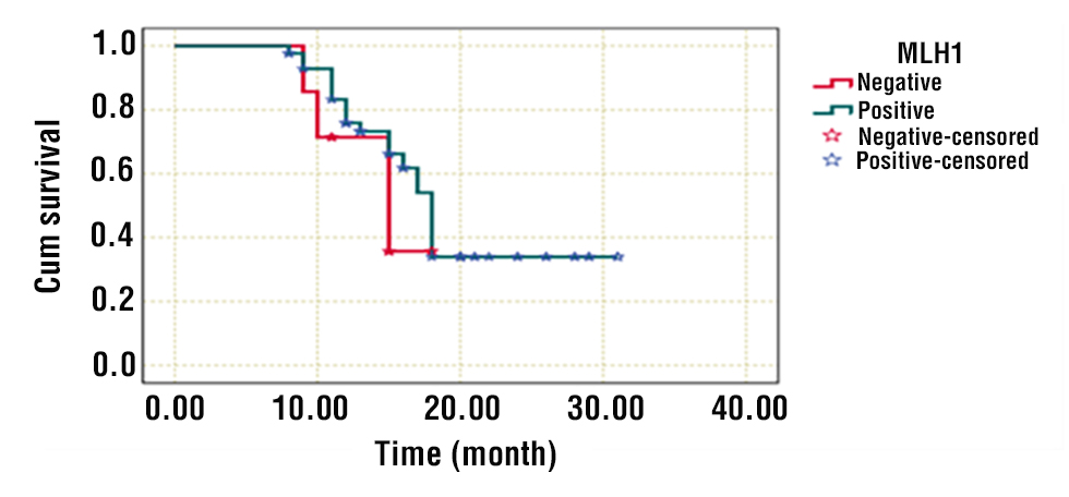
Figure 3 - (a, b) The loss of MLH1 protein expression in colon and rectal cancer tissues (red arrow); (c, d) Positive expression of MLH1 protein in colon and rectal cancer tissues (green arrow).
Discussion
The study results showed that the mean age of patients with CRC was 55 years . These findings are consistent with those reported in Baghdad (55 years) (10), Iran (56 years) (11), and Jordan (53 years) (12). However, they are inconsistent with the findings of a study conducted in Poland, which reported a higher mean age at diagnosis of 65 years (13). Overall, a mean age of 55 years at diagnosis may be considered relatively early on a global scale, particularly when compared with some Arab and Western populations Differences in the age at diagnosis between populations may reflect variations in demographic characteristics, genetic background, environmental exposures, and lifestyle factors (14).
The results indicated a 14% Loss of MLH1 expression among colorectal cancer cases, and loss of MLH1 expression has been reported to be associated with epigenetic silencing of the MLH1 gene in colo-rectal cancer (15,16). The observed frequency in the present study is consistent with previous reports of comparable rates, including 13% (17), 12% (18), and 7.7% (19), whereas a higher rate of 22% was reported in another study (20).
The results also indicated that MLH1 loss was more prevalent among patients aged < 50 years. These findings are consistent with those of previous studies (21,22), which reported a higher frequency of MLH1 loss of expression in younger age groups. In contrast, other studies (23,24) have demonstrated that MLH1 loss of expression is more common in patients older than 50 years. The increasing incidence of colorectal cancer among younger individuals has been reported in several populations worldwide; however, the factors underlying this trend remain incompletely understood (25).
In terms of sex, a higher frequency of MLH1 expression was observed in males. These findings are consistent with those of previous studies (26,27), which reported a greater proportion of MLH1 loss of expression in males than in females. In contrast, other studies (28,29) have demonstrated that the loss of expression is more frequent in females. The association between sex and MLH1 expression status remains inconsistent across studies, suggesting that multiple factors may influence this relationship (30).
Regarding family history, the majority of MLH1 loss cases were observed in patients without a positive family history. This observation is in agreement with the predominance of sporadic colorectal cancer cases reported in the literature. Most colorectal cancers occur sporadically, whereas a smaller proportion are associated with hereditary syndromes involving mismatch repair deficiency (31). These findings are consistent with those reported in (32), which demonstrated no significant association between protein expression status and family history.
With respect to histopathological variables and MLH1 protein expression status, the results showed that the highest number of MLH1 loss cases occurred in conventional adenocarcinomas. These findings are consistent with those reported previously (33,34), which demonstrated that the majority of MLH1 loss cases were observed in conventional adenocarcinoma.
The frequency of MLH1 protein loss is higher in adenocarcinomas. Previous studies have reported an association between MMR deficiency, including MLH1 loss, and mucinous histology in colorectal cancer (35,36).
In terms of tumor location, MLH1 expression was more prevalent in right-sided colon tumors. Right-sided colorectal cancers have been reported to exhibit distinct molecular characteristics, including a higher frequency of MMR deficiency and microsatellite instability, whereas left-sided tumors are more commonly associated with chromosomal instability and alterations in genes such as TP53 and KRAS (37). These findings are consistent with those reported in (34), which demonstrated a higher prevalence of MLH1 loss in right-sided tumors but differed from the findings of (38), which indicated that loss of expression was more common in left-sided colon tumors.
In terms of tumor differentiation, the results indicated that all cases of MLH1 expression loss were observed in moderately differentiated tumors. This finding is consistent with the distribution of tumor grades in the present study. Similar results were reported by (34), who found that most cases of MLH1 loss occurred in moderately differentiated tumors. In contrast, other studies (39) have reported a higher frequency of MLH1 loss in poorly differentiated tumors.With respect to tumor stage, MLH1 loss of expression was predominantly observed in stage T3 and T4 tumors. Similar findings were reported by (41), who demonstrated that the majority of MLH1 loss cases occurred in T3 and T4 tumors. The predominance of advanced tumor stage in the present cohort should be considered when interpreting these findings (40).
In lymphatic metastasis, MLH1 expression was predominantly observed in tumors without lymph node metastasis (N0). These findings are consistent with those reported in (34), which demonstrated that most cases of MLH1 loss occurred in the non-metastasized group. Previous studies have reported an association between MMR deficiency and reduced lymph node involvement in a subset of colorectal cancers (42). In terms of disease-free survival, no significant difference was observed between patients with positive MLH1 expression and those with loss of expression. These findings are consistent with those reported in (43), which similarly found no statistically significant difference in survival among colorectal cancer patients according to MLH1 expression status.
Conclusion
The majority of colorectal cancer cases occurred in older patients and males, with 14% exhibiting loss of MLH1 protein expression. A higher rate of MLH1 loss was observed in patients younger than 50 years, conventional adenocarcinoma, right-sided colon tumors, and stageT3 tumors. No significant difference in overall survival was detected between patients with MLH1 expression and those with loss of expression.
Authors’ Contributions
All researchers collaborated continuously from the initial preparation of the research plan and the finalization of the manuscript title, through sample collection, and finally to the completion of all research requirements. The authors have no conflicts of interest to declare.
Acknowledgments
The authors express their thanks and appreciation to the Histopathology Laboratory at Al-Hussein Teaching Hospital and the Health Department of thi-Qar Governorate / Iraq for their assistance in providing information and data related to our current study.
Conflicts of Interest
The authors declare no conflict of interest regarding this article.
Funding: None.
References
1. Huber AR, Pleguezuelos-Manzano C, Puschhof J, Ubels J, Boot C, Saftien A, et al. Improved detection of colibactin-induced mutations in genotoxic E. coli in organoids and colorectal cancer. Cancer Cell. 2024;42(3):487-496.
2. Luo H, Cui L, Shen K, Li R, Wang Z, Xie Z. HER2 Overexpression and Mismatch Repair Deficiency are Correlated with Malignancy in Colorectal Cancer. Cancer Manag Res. 2021;13:3443-3454.
3. Arends JW. Molecular interactions in the Vogelstein model of colorectal carcinoma. J Pathol. 2000;190(4):412-416.
4. Do Nascimento DJMB. Mutação BRAF V600E na história natural do cancro colorretal: implicações prognósticas e terapêuticas [doctoral dissertation]. Coimbra: Universidade de Coimbra; 2024.
5. Kasi A, Handa S, Bhatti S, Umar S, Bansal A, Sun W. Molecular pathogenesis and classification of colorectal carcinoma. Curr Colorectal Cancer Rep. 2020;16:97-106.
6. Sinicrope FA and Sargent DJ. Molecular pathways: microsatellite instability in colorectal cancer: Prognostic, predictive, and therapeutic implications. Clin Cancer Res. 2012;18(6):1506-1512.
7. Boland CR and Goel A. Microsatellite instability in colorectal cancer. Gastroenterology. 2010;138(6):2073-2087.
8. Lynch HT, Snyder CL, Shaw TG, Heinen CD, Hitchins MP. Milestones of Lynch syndrome: 1895–2015. Nat Rev Cancer. 2015;15(3):181-194.
9. Rodríguez–Soler M, Pérez–Carbonell L, Guarinos C, Zapater P, Castillejo A, Barberá VM, et al. Risk of cancer in cases of suspected lynch syndrome without germline mutations. Gastroenterology. 2013;144(5):926-932.
10. Zubairi RB, Hasan SA and Okab HF. Effects of IL-17 and IL-23 levels in women with Polycystic Ovary Syndrome. Reprod Heal East Eur. 2025;15(1):43–50.
11. Farhad RM, Saleh ES, Alsammarraie AZ. Clinicopathological features of colorectal cancer in the Iraqi population focusing on age and early onset of malignancy: A descriptive cross-sectional study. Al-Rafidain J Med Sci. 2023;5:86-91.
12. Elgohary SA, Abdelsalam SM, Khaled RST. Correlation between tumor location, clinicopathological characteristics, and mismatch repair protein (MMR) Status of Colorectal Cancer In Egyptian Patients. Egypt J Pathol. 2023;43(2):127-136.
13. Awad H, Elshebli S, Hasan K, Eid Y, Obeidat F, Alzyoud M, et al. Comparison of Clinicopathological and Immunohistochemical Features of Colorectal Carcinoma between Young and Old Age Groups. Diagnostics. 2024;14(16):1743.
14. Lewandowska A, Rudzki G, Lewandowski T, Stryjkowska-Gora A, Rudzki S. Risk factors for the diagnosis of colorectal cancer. Cancer Control. 2022;29:10732748211056692.
15. Syed AR, Thakkar P, Horne ZD, Abdul-Baki H, Kochhar G, Farah K, et al. Old vs. new: risk factors predicting early onset colorectal cancer. World J Gastrointest. Oncol. 2019;11(11):1011-1020.
16. Grady WM, Markowitz SD. Molecular pathogenesis of colorectal cancer and its potential application to colorectal cancer screening. Dig Dis Sci. 2015;60(3):762-772.
17. Goshayeshi L, Ghaffarzadegan K, Khooei A, Esmaeilzadeh A, Khorram MR, Mozaffari HM, et al. Prevalence and clinicopathological characteristics of mismatch repair-deficient colorectal carcinoma in early onset cases as compared with late-onset cases: A retrospective cross-sectional study in Northeastern Iran. BMJ Open. 2018;8(8):e023102.
18. Vlahović I, Rajc J, Švagelj I, Šolić K, Švagelj D.: Potential predictors for CDX2 expression loss and mismatch repair deficiency in colo-rectal cancer. Pathol Oncol Res. 2023;29:1610908.
19. Sayar I, Akbas EM, Isik A, Gokce A, Peker K, Demirtas L, et al. Relationship between mismatch repair deficiency, CDX2 loss, p53, and E-cadherin in colon carcinoma, and suitability of using a double panel of mismatch repair proteins by immunohistochemistry. Pol J Pathol. 2015;66(3):246-253.
20. Maraqa B, Al-Shbool G, Abu-Shawer O, Souleiman M, Alshakhatreh O, Al-Omari A, et al. Frequency of mismatch repair protein (MMRP) deficiency among young jordanians diagnosed with Colorectal Carcinoma (CRC). Gastroenterol Res. Pract. 2020;2020:5632984.
21. Bai H, Wang R, Cheng W, Shen Y, Li H, Xia W, et al. Evaluation of concordance between deficient mismatch repair and microsatellite instability testing, and their association with clinicopathological
features in colorectal cancer. Cancer Manag Res. 2020;12:2863-2873.
22. Manta BA, Ilie AC, Marc F, Nistor D, Mazilu PO, Borza C. Clinical Molecular Immunohistochemistry Mismatch Repair Mutations in Lynch Syndrome in Patients Under 50 Years: A Systematic Review. Biomedicines. 2025;13(5):1062.
23. Melincovici CS, Boşca AB, Şuşman S, Cutaş A, Mărginean M, Ilea A, et al. Assessment of mismatch repair deficiency, CDX2, beta-catenin and E-cadherin expression in colon cancer: molecular characteristics and impact on prognosis and survival–an immunohistochemical study. Rom J Morphol Embryol. 2021;61(3):715-725.
24. Soliman NA, Morsia DF, Helmy NA. Immunohistochemical expression of MMR proteins with clinicopathological correlation in colorectal cancer in Egypt. Open Access Maced J Med Sci. 2019;7(10):1608-1614.
25. Hashmi AA, Bukhari U, Rizwan R, Faisal F, Kumar R, Malik UA, et al. Mismatch Repair Deficient (dMMR) Colorectal Carcinoma in a Pakistani Cohort: Association With Clinical and Pathological Parameters. Cureus. 2023;15(8):e43242.
26. Durhuus JA, Therkildsen C, Kallemose T, Nilbert M. Colorectal cancer in adolescents and young adults with Lynch syndrome: a Danish register-based study. BMJ Open. 2021;11(12):e053538.
27. Bhattarai M, Juhari WKW, Lama R, Pun CB, Yusof W, Rahman WFA, et al. MLH1 and MSH2 mismatch repair protein profile using immunohistochemistry in Nepalese colorectal cancer patients. Med J Indones. 2020;29(2):183-189.
28. Guo L, Yu Z, Li Q, Liang X, Yang L. Correlation of MLH1 and MSH2 levels with clinicopathologic characteristics in colorectal cancer. Am J Transl Res. 2023;15(2):1107-1115.
29. Mao J, He Y, Chu J, Hu B, Yao Y, Yan Q, et al. Analysis of clinical characteristics of mismatch repair status in colorectal cancer: a multicenter retrospective study. Int J Colorectal Dis. 2024;39(1):100.
30. Haghighatdoost F, Mehrabani-Zeinabad K, Hajihashemi P, Mohammadifard N, Adibi P. Burden of colorectal cancer and its risk factors in the North Africa and Middle East (NAME) region, 1990–2019: a systematic analysis of the global burden of disease study. BMC Public Health. 2024;24(1):557.
31. Rico-Méndez MA, Trujillo-Rojas MA, Ayala-Madrigal MDL, Hernández-Sandoval JA, González-Mercado A, Gutiérrez-Angulo M, et al. MLH1 methylation status and microsatellite instability in patients with colorectal cancer. Genes. 2025;16(2):182.
32. Liu Y, Guo YH, Luo Y, Sun L, Zhao S, Shao B, et al. Mismatch repair protein expression of colorectal cancer: a retrospective analysis of 3 428 cases. Zhonghua Bing Li Xue Za Zhi. 2021;50(4):369-375.
33. Tanaka H, Deng G, Matsuzaki K, Kakar S, Kim GE, Miura S, et al. BRAF mutation, CpG island methylator phenotype and micro-satellite instability occur more frequently and concordantly in mucinous than non‐mucinous colorectal cancer. Int J Cancer. 2006;118(11):2765-2771.
34. Sun CY, Chiang JM, Chen TC, Hung HY, You JF. Different surgical outcome and follow-up status between dMMR and pMMR colorectal cancer patients who fulfilled with Amsterdam-II criteria. World J Surg Oncol. 2020;18(1):195.
35. Andrici J, Farzin M, Sioson L, Clarkson A, Watson N, Toon CW, et al. Mismatch repair deficiency as a prognostic factor in mucinous colorectal cancer. Mod Pathol. 2016;29(3):266-274.
36. Mik M, Berut M, Dziki L, Trzcinski R, Dziki A. Right-and left-sided colon cancer–clinical and pathological differences of the disease entity in one organ. Arch Med Sci. 2017;13(1):157-162.
37. Idrissi MB, El Bouhaddouti H, Mouaqit O, Ousadden A, Ait Taleb K, Benjelloun EB. Left-Sided Colon Cancer and Right-Sided Colon Cancer: Are They the Same Cancer or Two Different Entities? Cureus. 2023;15(4):e38312.
38. Ionescu VA, Gheorghe G, Baban IA, Barbu A, Antonie NI, Georgescu TF, et al. Clinical, Immunohistochemical, and Inflammatory Profiles in Colorectal Cancer: The Impact of MMR Deficiency. Diagnostics. 2025;15(17):2141.
39. Wang SM, Jiang B, Deng Y, Huang SL, Fang MZ, Wang Y. Clinical significance of MLH1/MSH2 for stage II/III sporadic colorectal cancer. World J Gastrointest Oncol. 2019;11(11):1065-1080.
40. Tra ĐT, Thịnh PV, Dũng TN. Study on microsatellite instability in colorectal adenocarcinoma using immunohistochemistry. J 108-Clin Med Pharm. 2023;18(2):141-148.
41. Belt ET, Te Velde EA, Krijgsman O, Brosens RPM, Tijssen M, Van Essen HF, et al. High lymph node yield is related to microsatellite instability in colon cancer. Ann Surg Oncol. 2012;19(4):1222-1230.
42. Shin US, Cho SS, Moon SM, Park SH, Jee SH, Jung EJ, et al. Is microsatellite instability really a good prognostic factor of colorectal cancer? Ann Coloproctol. 2014;30(1):28-34.
43. Denčić T, Krstić M, Petrović A, Jovičić-Milentijević M, Radenković G, Jović M, et al. Prognostic value and immunohistochemical analysis of mismatch repair deficiency in patients with stage II and III colorectal carcinoma - A single-center study. Medicina. 2020; 56(12):676.
Full Text Sources:

Abstract:

Views: 39


For Authors
Journal Subscriptions

Jun 2026
Supplements
Instructions for authors
Online submission
Contact
e-ISSN: 2601 - 1700 (online)
ISSN-L: 2559 - 723X
Journal Abbreviation: Surg. Gastroenterol. Oncol.
Surgery, Gastroenterology and Oncology (SGO) is indexed in:
- SCOPUS
- EBSCO
- DOI/Crossref
- Google Scholar
- SCImago
- Harvard Library
- Open Academic Journals Index (OAJI)
Surgery, Gastroenterology and Oncology (SGO) is an open-access, peer-reviewed online journal published by Celsius Publishing House. The journal allows readers to read, download, copy, distribute, print, search, or link to the full text of its articles.
Time to first editorial decision: 25 days
Rejection rate: 61%
CiteScore: 0.2
 IASGO Society News

Meetings and Courses in 2025
Meetings and Courses in 2024
Meetings and Courses in 2023
Meetings and Courses in 2022
Meetings and Courses in 2021
Meetings and Courses in 2020
Meetings and Courses in 2019
Verona expert meeting 2019

Surgery, Gastroenterology and Oncology applies the Creative Commons Attribution Non Commercial (CC BY-NC 4.0) license, which permits readers to copy and redistribute the material in any medium or format, remix, adapt, build upon the published works non-commercially, and license the derivative works on different terms, provided the original material is properly cited and the use is non-commercial. Please see: https://creativecommons.org/licenses/by-nc/4.0/
Publisher’s Note:
The opinions, statements, and data contained in article are solely those of the authors and not of Surgery, Gastroenterology and Oncology journal or the editors. Publisher and the editors disclaim responsibility for any damage resulting from any ideas, instructions, methods, or products referred to in the content.