Surgery, Gastroenterology and Oncology
|
|
Objective: This study aims to evaluate of some inflammatory markers, oxidative stress indicators, and vitamins C and E levels in patients with gastric ulcer, and to compare these markers between those infected and non-infected with H. pylori.
Methods: The study initially evaluated 320 participants for demographic analysis. Laboratory and biochemical assessments were performed on a strictly selected sub-sample of 88 eligible participants divided into three groups: Gastric Ulcer only (n=34), Gastric Ulcer with H. pylori (n=34), and a healthy Control group (n=20). Clinical diagnosis was confirmed using esophagogastroduodenoscopy (EGD) and H. pylori testing. To eliminate physiological bias, statistical analyses were stratified by gender.
Results: The results revealed a significant increase in the levels of inflammatory cytokines such as IL-18, IFN, and (CRP) among patients, particularly when the ulcer was associated with H. pylori infection, in both sexes. Furthermore, the results showed a severe imbalance in oxidative stress, characterized by elevated levels of MDA against a decrease in the activity of antioxidant enzymes. Regarding vitamins, no significant differences were observed in males, while vitamin E was significantly decreased in females with gastric ulcer only (without H. pylori infection).
Conclusion: H. pylori infection leads to physiological dysfunction manifested by the stimulation of an inflammatory response and elevated levels of oxidative stress, reflecting the impact of the interaction between gender and the nature of the disease on inflammatory pathways.
Keywords: gastric ulcer, Helicobacter pylori, oxidative stress, inflammatory markers, cytokines
INTRODUCTION
Gastric ulcer is a highly prevalent gastrointestinal disorder characterized by localized erosion of the gastric mucosal lining, resulting from an imbalance between aggressive factors (e.g., gastric acid, pepsin) and mucosal protective mechanisms (1,2). Historically attributed to psychological stress and poor diet (3), the discovery of H. pylori revolutionized our understanding, establishing it as a primary etiological agent (4,5).
Beyond H. pylori, excessive use of (NSAIDs) is a major contributing factor. NSAIDs inhibit cyclooxygenase enzymes, reducing protective prostaglandin synthesis, thus weakening the mucosal barrier and increasing susceptibility to acid-induced injury (6,7). Additional risk factors include psychological stress, smoking, genetic predisposition, and poor dietary habits (8,9). Clinically, gastric ulcers typically present with epigastric burning pain, often worsening on an empty stomach, and may progress to serious complications such as hemorrhage or perforation if untreated (10).
Recent research has increasingly focused on the role of inflammatory and oxidative stress biomarkers in disease progression. H. pylori infection induces excessive production of ROS, leading to elevated intracellular oxidative stress, lipid peroxidation, and activation of inflammatory pathways (11). Key inflammatory cytokines such as IL-18, IFN, and IL-36 are central to the host immune response (12,13). Concurrently, oxidative stress markers like MDA and antioxidant enzymes (SOD, CAT, GPX) reflect the extent of cellular damage and defense depletion (14,15).
Furthermore, emerging evidence highlights sex-based differences in immune and oxidative responses, potentially mediated by sex hormones such as estrogen (16,17). Therefore, assessing these biomarkers in both males and females is crucial for a comprehensive understanding of disease pathophysiology.
Based on the foregoing data, the present study was undertaken to evaluate and analyze selected indicators of inflammation and oxidative stress in patients with gastric ulcer in the district of Al-Rifai, with a comparative approach between cases associated with H. pylori infection and those without. The current study aimed to elucidate the extent to which this infection contributes to the exacerbation of the inflammatory response and the escalation of oxidative stress severity, thereby facilitating the development of more accurate diagnostic and therapeutic insights for patients in the region.
METHODS AND MATERIALS
Study Design
The study included 320 participants of both sexes. A total of 300 gastric ulcer patients were enrolled for demographic data collection and analysis. Laboratory and biochemical evaluations were conducted on a strictly selected sub-sample of 88 eligible participants divided into three groups: Gastric Ulcer only (n=34), Gastric Ulcer with H. pylori (n=34), and a healthy Control group (n=20). Diagnoses were confirmed via endoscopy (EGD) and H. pylori testing. To eliminate sex-based physiological bias, statistical analyses were conducted separately for each gender.
Inclusion and Exclusion Criteria
Eligible participants for the laboratory subsample were adults aged 18-60 years with clinically and endoscopically confirmed gastric ulcers (with or without H. pylori infection) or healthy controls who provided informed consent. Exclusion criteria comprised individuals with chronic liver, kidney, cardiovascular, or metabolic diseases; gastrointestinal malignancies; previous gastric surgery; active smoking or alcohol consumption; or recent use (within the past month) of NSAIDs, cortico-steroids, immunosuppressants, anti-oxidant supplements, or H. pylori eradication therapy to prevent baseline biomarker confounding.
Clinical Diagnosis of Gastric Ulcer and Helicobacter pylori Status Assessment
Clinical diagnosis of gastric ulcers was established and confirmed by specialized gastroenterologists using upper gastrointestinal endoscopy (esophago-gastroduodenoscopy, EGD). To evaluate H. pylori infection status, a rapid lateral flow immunochromato-graphic assay based on a double-antibody sandwich technique was performed for the qualitative detection of H. pylori antigen in fresh stool specimens. Fresh stool (50–100 mg) was collected from three distinct areas of the sample, homogenized in the provided extraction buffer, shaken vigorously for 15–20 seconds, and allowed to settle for 2 minutes. Three to four drops of the supernatant were dispensed into the sample well of the cassette and incubated at room temperature. Results were visually interpreted at 15 minutes: the appearance of both the control (C) and test (T) bands indicated a positive result, whereas a single control (C) band indicated a negative result. Samples exhibiting no control band were deemed invalid and retested. Patients were subsequently categorized based on concordant endoscopic findings and stool antigen test results.
Confounding Variables Control and Justification for Sex-Stratified Analysis
Potential confounding variables (such as age, underlying chronic comorbidities, medication use including NSAIDs and antioxidants, and smoking status) were controlled a priori through stringent inclusion and exclusion criteria and balanced sex-matching across the biochemical sub-groups.
Statistical analyses were intentionally stratified by sex rather than pooled to account for well-documented biological dimorphism. Estrogen and sex-specific hormonal profiles significantly modulate immune responses, cytokine expression (e.g., IL-18 and IL-36), and baseline antioxidant enzyme activities (SOD, CAT, GPX). Combining male and female data would introduce substantial physiological confounding and obscure sex-specific pathological mechanisms induced by H. pylori infection and gastric ulceration.
Laboratory Measurements and Studied Parameters
This study was conducted in accordance with the principles of the Declaration of Helsinki, with official ethical approval obtained from the Research Committee of the Dhi‑Qar Health Directorate (Ref No: 228/2025); written informed consent was secured from all participants, and the confidentiality of their data was strictly maintained. The parameters investigated included inflammatory markers, namely CRP, IFN, and interleukins (IL‑18 and IL‑36); oxidative stress parameters, including MDA, LPO, and the enzymatic antioxidant activities of SOD, CAT, and GPX; as well as the levels of vitamin C and vitamin E. All laboratory measurements were performed using commercially available (ELISA) kits supplied by Cloud‑Clone Corp. (Wuhan, China), strictly adhering to the operating protocols provided by the manufacturer.
Statistical Analysis
Data were analyzed using SPSS (version 27) and are expressed as mean ± SD. Differences among groups were assessed using one-way ANOVA followed by Tukey’s HSD post-hoc test to correct for multiple comparisons. Analyses were stratified by sex to eliminate physiological bias. Potential confounders were controlled via strict selection criteria. Post-hoc power analysis (G*Power v3.1) confirmed that the sub-sample (n=88) achieved a statistical power >80% at p ≤ 0.05.
RESULTS
Relationship Between Gastric Ulcer and Inflammatory Markers
Tables 1 and 2 illustrate the relationship between gastric ulcer and inflammatory markers for males and females, respectively, at a probability level of p ≤ 0.05. The results presented in table (1) reveal a significant increase in the concentrations of IL-18, IFN, and CRP, along with a significant decrease in the IL-36 concentration, in both the gastric ulcer group (Group 1) and the gastric ulcer group associated with H. pylori infection (Group 2), when compared to the healthy control group (Group 3), at a probability level of p ≤ 0.05.
Table 1 - the relationship between gastric ulcer and inflammatory markers in males
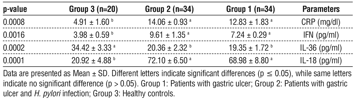
Table 2 - the relationship between gastric ulcer and inflammatory markers in females
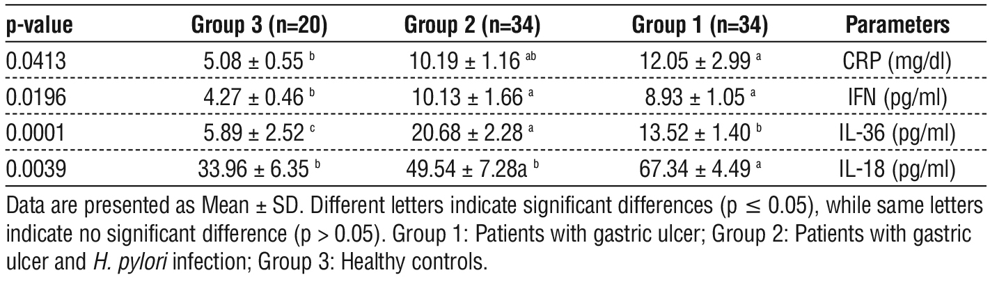
Furthermore, the results in table 2 indicated a significant increase in the concentrations of IL-18 and (CRP) in Group 1 (gastric ulcer) compared to Group 3 (healthy controls). The findings also demonstrated a significant increase in IFN concentration in both Group 1 and Group 2 relative to Group 3. Additionally, the results revealed a significant increase in IL-36 concentration in both Group 1 and Group 2 compared to Group 3, with Group 2 showing a significant increase compared to Group 1, at the aforementioned probability level.
Relationship Between Gastric Ulcer and Oxidative Stress
The results presented in table 3 revealed the relationship between gastric ulcer and oxidative stress in males. A significant increase in MDA concentration was observed in both Group 1 (gastric ulcer) and Group 2 (gastric ulcer with H. pylori infection) when compared to Group 3 (healthy controls) at a probability level of p ≤ 0.05. Furthermore, the findings revealed a significant decrease in (GPX) concentration in Group 2 relative to Group 3, in addition to a significant increase in (CAT) concentration in both Group 1 and Group 2 compared to Group 3 at the aforementioned probability level. On the other hand, the results indicated a significant increase in SOD concentration in Group 2 when compared to Group 3, and a significant decrease in LPO concentration in Group 2 relative to Group 3, as shown in table 3.
Table 3 - the relationship between gastric ulcer and oxidative stress in males
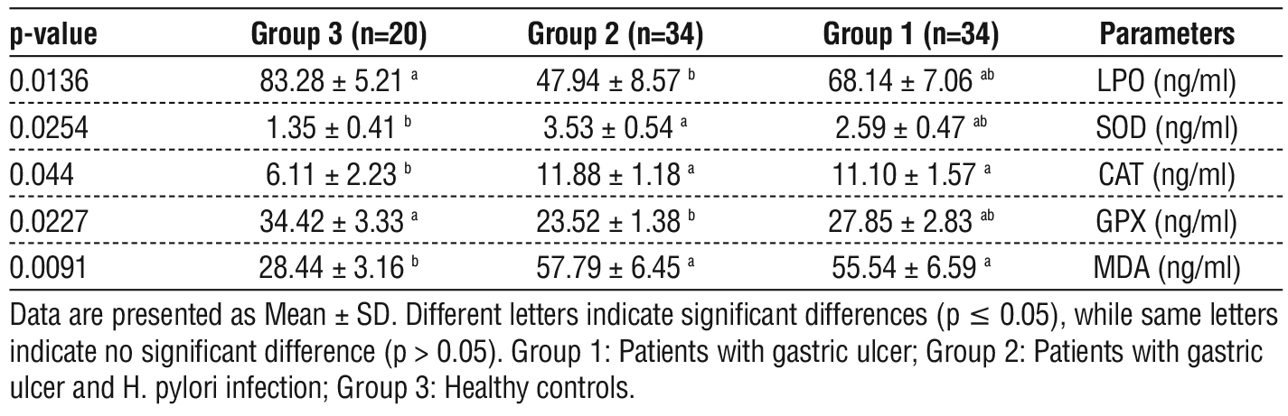
Table 4 - the relationship between gastric ulcer and oxidative stress in females
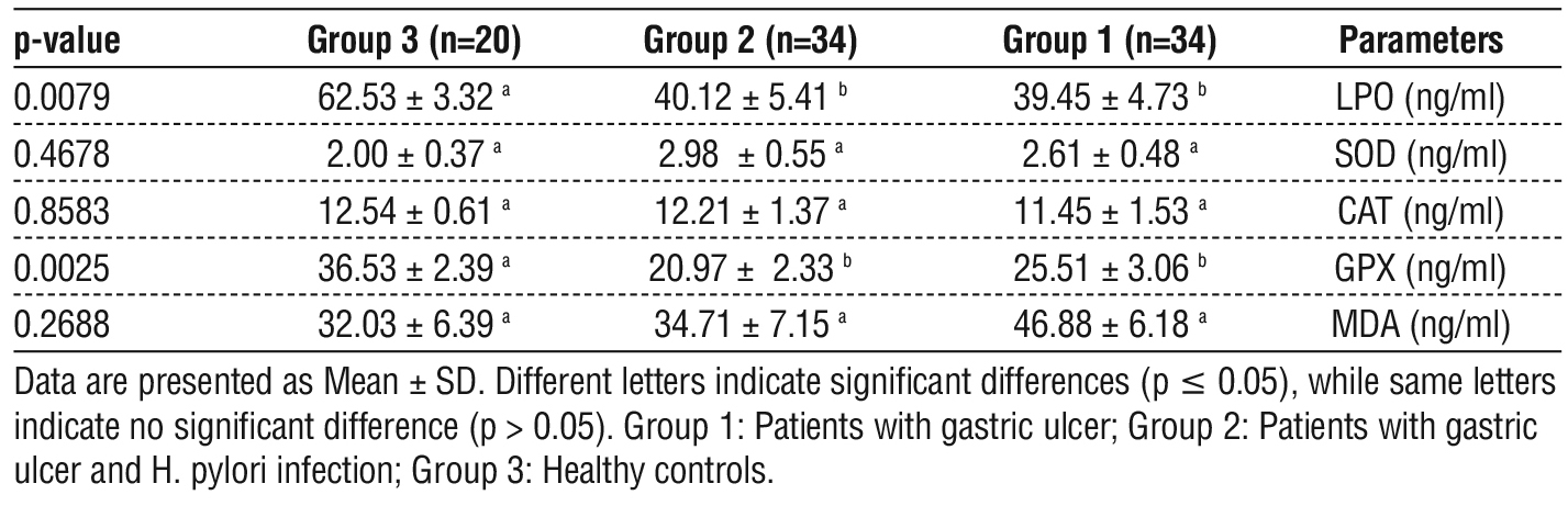
On the other hand, the results presented in table 4 showed the relationship between gastric ulcer and oxidative stress in females. It is observed that there is no significant difference in the concentrations of MDA, CAT, and SOD among the three study groups at a probability level of p ≤ 0.05. However, a significant decrease in the concentrations of both GPX and LPO was observed in Group 1 and Group 2 when compared to Group 3 at the aforementioned probability level.
Relationship Between Gastric Ulcer and Vitamins
Table 5 appeared the relationship between gastric ulcer and serum levels of certain vitamins in males. The results indicate no significant differences between the groups of patients with gastric ulcer and those with H. pylori-associated ulcer (Groups 1 and 2) compared to the healthy control group (Group 3) at a probability level of p ≤ 0.05.
Table 5 - the relationship between gastric ulcer and certain vitamins in males

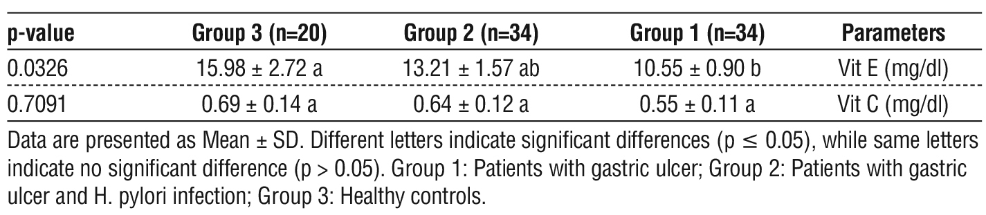
Table 6 shows the relationship between gastric ulcer and serum levels of certain vitamins in females at a probability level of p ≤ 0.05. The results reveal no significant differences in vitamin C levels among all groups. However, a significant decrease in vitamin E levels was observed in the first group (patients with gastric ulcer) compared to the third group (healthy controls), which did not differ significantly from the second group (patients with gastric ulcer and H. pylori infection).
Table 6 - the relationship between gastric ulcer and certain vitamins in females
DISCUSSION
The present study investigated the relationship between gastric ulcer, H. pylori infection, and key inflammatory and oxidative stress biomarkers, revealing significant physiological disturbances with clear sex-based differences.
The observed alterations in circulating inflammatory cytokines and redox parameters provide direct systemic evidence of the mucosal damage occurring during gastric ulceration and H. pylori infection. Specifically, the significant elevations in serum IL-18, IFN, and CRP directly reflect an activated Th1-mediated inflammatory state and systemic immune response in infected patients. While the activation of intracellular pathways - such as NOD1/caspase-1 pyroptosis or downstream mucosal gene expressions - represents plausible, literature-supported mechanistic frameworks for these observations, our serum-based findings directly demonstrate the functional end-products of this immune activation. Similarly, the marked increase in lipid peroxidation (MDA) concurrent with depleted antioxidant enzyme capacity (GPX) directly confirms systemic redox imbalance, directly reflecting the extent of oxidative tissue injury induced by the infection and mucosal lesions.
Regarding inflammatory markers, the significant elevation of IL-18 and IFN in both patient groups (tables 1 & 2) strongly correlates with the immune response triggered by H. pylori. The bacterium activates the NOD1/caspase-1 pathway, promoting IL-18 processing through pyroptosis, which sub-sequently recruits neutrophils and macrophages, exacerbating tissue injury (18). The elevated IFN levels further indicate a predominant Th1 response, consistent with the immunopathology of H. pylori-associated ulcers (19,20).
Concurrently, regarding IL-36, the significant decrease observed in males (table 1) alongside its marked elevation in female patients (table 2) highlights a sex-specific mucosal immune response. While the reduction in males may indicate a potential dysfunction or excessive consumption of the tissue-repair axis during chronic inflammation, the heightened expression in females suggests enhanced mucosal immune activation and a differential regulatory role modulated by sex-specific hormonal profiles (21,22).
Regarding oxidative stress, our findings indicate a severe imbalance in ulcer patients. The significant
elevation of MDA (table 3) confirms enhanced LPO due to excessive free radicals released by infiltrating neutrophils during gastric inflammation (14). This is supported by the observed decrease in GPX and LPO levels in H. pylori-infected patients (Group 2), attributed to the exhaustive consumption of these antioxidants in a futile attempt to counteract the overwhelming oxidative burden (15,23). However, the significant increase in SOD and CAT activities in males with H. pylori infection suggests a compensatory upregulation of the antioxidant defense system. This is consistent with studies showing that the body attempts to enhance enzymatic gene expression to protect the mucosa and facilitate healing (24). Nevertheless, persistent infection sustains chronic inflammation, preventing a complete restoration of redox balance and maintaining elevated MDA levels despite increased SOD/CAT activity (25). In females, the less pronounced changes in MDA, SOD, and CAT compared to males may reflect protective hormonal effects, such as estrogen's antioxidant properties, which modulate oxidative stress differently between sexes (16,17). The significant decline in GPX and LPO in both female patient groups reinforces the vulnerability of the glutathione-dependent antioxidant system under chronic inflammatory stress (23).
Regarding vitamin levels, the absence of significant differences in males and vitamin C levels in all groups suggests that dietary intake or malabsorption may not be the primary driver in this population. However, the significant decrease in serum vitamin E in females with gastric ulcer alone (Group 1) may reflect its specific depletion as a major lipophilic antioxidant combating lipid peroxidation within the inflamed gastric mucosa (26-27). This reduction could also result from chronic inflammation-induced oxidative consumption rather than the mere presence of infection, as Group 2 (with H. pylori) showed intermediate levels without statistical significance.
Clinical Significance and Practical Applications of Evaluated Biomarkers
Beyond their pathophysiological involvement, the evaluated biomarkers offer distinct clinical potential for managing gastric ulcer disease and H. pylori infection. Specifically, tracking inflammatory cytokines (such as IL-18 and IL-36) alongside systemic oxidative stress markers (e.g., MDA and GPX activity) provides a non-invasive tool to monitor disease severity and mucosal healing status without relying solely on repeated endoscopies. Clinically, profound elevations in lipid peroxidation (MDA) concurrent with depleted antioxidant defenses signal severe mucosal injury, which could identify high-risk patients prone to ulcer complications such as bleeding or recurrence. Furthermore, sex-stratified biomarker profiles suggest that therapeutic strategies - particularly antioxidant supplementation and anti-inflammatory adjunct therapies - should be tailored individually according to patient sex to optimize clinical outcomes and restore redox homeostasis.
Study Limitations
This study has several limitations. First, the laboratory sub-sample size (n=88) was relatively small, though statistically sufficient according to power analysis. Second, the cross-sectional design prevents establishing causal relationships over time. Third, H. pylori status was evaluated via stool antigen testing and endoscopy without histopathological grading or bacterial strain virulence profiling (e.g., CagA/VacA). Finally, despite strict exclusion criteria, unmeasured environmental factors such as dietary variations might still exert minor effects on oxidative stress parameters.
Overall, these findings confirm that H. pylori infection acts as a potent trigger exacerbating both inflammatory responses and oxidative stress, surpassing the defensive capacity of the host's anti-oxidant systems. The clear sex-based differences observed underscore the critical interplay between
biological sex and disease pathology, which should be considered in future diagnostic and therapeutic
strategies. Further studies with larger sample sizes and longitudinal designs are recommended to validate these biomarkers as potential therapeutic targets.
CONCLUSIONS
Infection with H. pylori induces a profound physiological disturbance characterized by the activation of a systemic inflammatory response and the escalation of oxidative stress levels, accompanied by a marked depletion of antioxidant defenses. Furthermore, the nature of this immunological and biochemical response exhibits clear sex-based differences, reflecting a significant influence of the interaction between sex and pathology on inflammatory pathways.
Authors’ Contributions
Noor Al-Huda T. Basheet and Ali M. Hussein contributed equally to the study design, data colection, laboratory analyses and writing the manuscript. Both authors reviewed and approved the final version.
Conflicts of Interest
The authors declare no conflict of interest regarding this article.
Funding: None.
Ethics of Approval
This study was conducted in accordance with the principles of the Declaration of Helsinki, with official ethical approved obtained from the Research Committee of the Dhi - Qar Health Directorate (Ref No: 228/2025).
REFERENCES
1. Khan AH, Dar MA, Mir MA. Gastric ulcer: An overview. International Journal of Current Research in Physiology and Pharmacology. 2023; 7(3):1-7.
2. Erfan IA, Khodir AE, Ghattas M, Abo Elmaaty DM, Mesbah N, Wahba A. Gastric ulcer: An overview of pathophysiology, diagnosis, and management. Records of Pharmaceutical and Biomedical Sciences. 2025;9(1):162-186.
3. Cho JH, Jin SY. Diagnosis and treatment of Helicobacter pylori infection: past, present, and future. Future Microbiol. 2025;20(13): 913-929.
4. Malfertheiner P, Schulz C, Hunt RH. Helicobacter pylori Infection: A 40-year journey through shifting the paradigm to transforming the management. Dig Dis. 2024;42(4):299-308.
5. Crowe SE. Helicobacter pylori infection. N Engl J Med. 2019; 380(12):1158-1165.
6. Ko KA, Lee DK. Nonsteroidal anti-inflammatory drug-induced peptic ulcer disease. Korean J Helicobacter Up Gastrointest Res. 2025; 25(1):34-41.
7. Islam H, Siddiqui A, Islam R, Islam T, Ahmed S, Fahim M, et al. NSAID-induced gastric ulcer disease: a deleterious connection. Discov Med. 2024;36(188):1789-1799.
8. Guan Ningzi, Liu L. Microbial response to acid stress: Mechanisms and applications. Appl Microbiol Biotechnol. 2020;104(1):51-65.
9. Bailey JM. Gastrointestinal conditions: peptic ulcer disease. FP Essent. 2024;540:16-23.
10. Dimou F, Velanovich V. Perforations of the esophagus and stomach: what should I do? J Gastrointest Surg. 2015;19(2):400-6.
11. Suzuki H, Hirai M. Helicobacter pylori infection and oxidative stress. J Clin Biochem Nutr. 2024;75(3):178-182.
12. Buyukcolak-Cebeci Y, Timucin E, Akcelik-Deveci S, Mansur-Ozen N, Aydinlar T, Tiftikci A, et al. Determination of the Roles of H. pylori Outer Membrane Virulence Factors and Pyroptosis-Associated NLRP3, ASC, Caspase-1, Gasdermin D, IL-1b, and IL-18 in Ulcer and Gastritis Pathogenesis. Biology (Basel). 2025;14(6):634.
13. Lin Y, Liu K, Lu F, Zhai C, Cheng F. Programmed cell death in Helicobacter pylori infection and related gastric cancer. Front Cell Infect Microbiol. 2024;14:1416819.
14. Bhattacharyya A, Chattopadhyay R, Mitra S, Crowe SE. Oxidative stress: an essential factor in the pathogenesis of gastrointestinal mucosal diseases. Physiol Rev. 2014;94(2):329-54.
15. Butcher LD, den Hartog G, Ernst PB, Crowe SE. Oxidative stress resulting from Helicobacter pylori infection contributes to gastric carcinogenesis. Cell Mol Gastroenterol Hepatol. 2017;3(3):316-322.
16. Wilkinson NM, Chen HC, Lechner MG, Su MA. Sex differences in immunity. Annu Rev Immunol. 2022;40:75-94.
17. Peng C, Li X, Li Y, Xu X, Ouyang Y, Li N, et al. Sex-specific effects of gastrointestinal microbiome disruptions on Helicobacter pylori-induced gastric carcinogenesis in INS-GAS mice. Biol Sex Differ. 2025;16(1):15.
18. Tran LS, Ying L, D'Costa K, Wray-McCann G, Kerr G, Le L, et al. NOD1 mediates interleukin-18 processing in epithelial cells responding to Helicobacter pylori infection in mice. Nat Commun. 2023;14(1):3804.
19. Fan J, Zhu J, Xu H. Strategies of Helicobacter pylori in evading host innate and adaptive immunity: insights and prospects for therapeutic targeting. Front Cell Infect Microbiol. 2024;14:1342913.
20. Zhao X, Tong D, Ferrero RL. Interleukin-18 produced by gastric epithelial cells protects against pre-neoplastic lesions in Helicobacter pylori infection in mice. Genes Immun. 2024;25(4):346-347.
21. Ngo VL, Kuczma M, Maxim E, Denning TL. IL-36 cytokines and gut immunity. Immunology. 2021;163(2):145-154.
22. Shirani M, Shariati S, Bazdar M, Ghamnak SF, Moradi M, Shams Khozani R, et al. The immunopathogenesis of Helicobacter pylori-induced gastric cancer: A narrative review. Front Microbiol. 2024;15:1395403.
23. Kouitcheu Mabeku LB, Faujo Nintewoue GF, Nguefak LDT, Timba IN, Ngatcha G, Tagni SM, et al. Oxidative stress dynamic among Helicobacter pylori-positive patients with various stages of pre-malignant histologic lesions. Asian Pac J Cancer Prev. 2025; 26(10):3609-3618.
24. Kim S, Kim JM, Shim SH, Chang HI. Anthocyanins accelerate the healing of naproxen-induced gastric ulcer in rats by activating antioxidant enzymes via modulation of Nrf2. Journal of Functional Foods. 2014;7:569-579.
25. Kuna Lucija, Jakab J, Smolic R, Raguz-Lucic N, Vcev A, Smolic M. Peptic ulcer disease: a brief review of conventional therapy and herbal treatment options. J Clin Med. 2019;8(2):179.
26. Nair, Satheesh, Norkus, E. P., Hertan, H., Pitchumoni, C. S. Micronutrient antioxidants in gastric mucosa and serum in patients with gastritis and gastric ulcer: does Helicobacter pylori infection affect the mucosal levels?. J Clin Gastroenterol. 2000;30(4):381-5.
27. Lee BJ, Jihye K, Kim KH. Association of gastric and duodenal ulcers with anthropometry and nutrients: Korean National Health and Nutrition Examination Survey (Knhanes II-IV) 2001-2009. PLoS One. 2018;13(11):e0207373.
Full Text Sources:

Abstract:

Views: 47


For Authors
Journal Subscriptions

Jun 2026
Supplements
Instructions for authors
Online submission
Contact
e-ISSN: 2601 - 1700 (online)
ISSN-L: 2559 - 723X
Journal Abbreviation: Surg. Gastroenterol. Oncol.
Surgery, Gastroenterology and Oncology (SGO) is indexed in:
- SCOPUS
- EBSCO
- DOI/Crossref
- Google Scholar
- SCImago
- Harvard Library
- Open Academic Journals Index (OAJI)
Surgery, Gastroenterology and Oncology (SGO) is an open-access, peer-reviewed online journal published by Celsius Publishing House. The journal allows readers to read, download, copy, distribute, print, search, or link to the full text of its articles.
Time to first editorial decision: 25 days
Rejection rate: 61%
CiteScore: 0.2
 IASGO Society News

Meetings and Courses in 2025
Meetings and Courses in 2024
Meetings and Courses in 2023
Meetings and Courses in 2022
Meetings and Courses in 2021
Meetings and Courses in 2020
Meetings and Courses in 2019
Verona expert meeting 2019

Surgery, Gastroenterology and Oncology applies the Creative Commons Attribution Non Commercial (CC BY-NC 4.0) license, which permits readers to copy and redistribute the material in any medium or format, remix, adapt, build upon the published works non-commercially, and license the derivative works on different terms, provided the original material is properly cited and the use is non-commercial. Please see: https://creativecommons.org/licenses/by-nc/4.0/
Publisher’s Note:
The opinions, statements, and data contained in article are solely those of the authors and not of Surgery, Gastroenterology and Oncology journal or the editors. Publisher and the editors disclaim responsibility for any damage resulting from any ideas, instructions, methods, or products referred to in the content.