Surgery, Gastroenterology and Oncology
Background: Full-thickness rectal prolapse (FTRP) is commonly treated using rectopexy techniques. However, these methods mainly focus on fixing the rectum and do not restore the physiological support mechanism of the pelvic floor. Based on the dual pelvic support axis theory, full-thickness rectal prolapse is assumed to result from failure of the posterior pelvic support axis (PPSA). At Hanoi Medical University Hospital, this technique has been performed since January 2022. Objective: To evaluate the early outcomes of laparoscopic reconstruction of the posterior pelvic support axis in female patients with FTRP and to analyze its pathophysiological basis compared with existing techniques.
Methods: A retrospective study of 14 female patients with FTRP who underwent PPSA reconstruction using a synthetic mesh. Surgical, clinical, and functional parameters were evaluated.
Results: The mean operative time was 114.6 minutes, with minimal blood loss (46 mL). No recurrence was recorded. The rate of fecal incontinence decreased from 85.7% to 28.6%, and constipation decreased from 78.8% to 42.9%. Quality of life improved significantly.
Conclusion: Laparoscopic PPSA reconstruction is a safe and effective method that restores the physiological support mechanism of the pelvic floor rather than simply fixing the rectum, opening a new approach in the treatment of FTRP.
Keywords: rectal prolapse, PPSA, synthetic mesh, laparoscopic
INTRODUCTION
Full-thickness rectal prolapse is a debilitating condition characterized by the protrusion and descent of the entire rectal wall through the anal canal, a pathology intimately linked to global pelvic floor dysfunction (1-5). The estimated prevalence is less than 0.5% in the general population, primarily affecting older adults (6,8,9). It is significantly more common in women, accounting for 80–90% of cases, with a female-to-male ratio ranging from 6:1 to 10:1 (8,10). The most common age of onset is over 50, particularly among elderly women with weakened pelvic floor support (8,11-13).
Regarding pathogenesis, FTRP has long been viewed as the consequence of redundancy and intussusception of the rectum through the anal canal. This perspective led to the development of surgical strategies focused on anatomical restoration via rectopexy or rectal resection (8,10). Classic approaches, such as posterior rectopexy and Ventral Mesh Rectopexy (VMR), have demonstrated efficacy in reducing recurrence rates and improving functional outcomes (6,9,11). However, these methods remain essentially "single-organ" approaches, treating the rectum as an independent entity without fully considering the role of the overall pelvic floor support and suspension system (15,16).
Despite various technical refinements, limitations persist. Posterior rectopexy, involving extensive dissection of the posterior rectal space, can damage the autonomic nervous system, thereby increasing postoperative constipation rates (7-9). Conversely, VMR—with its advantage of preserving the posterior plane and autonomic nerves—improves defecatory function, particularly constipation (11-13). However, this technique primarily targets the anterior rectal wall and the rectovaginal septum, without directly addressing the underlying supporting structures, especially the perineal body (13,17). This may explain the persistence or incomplete improvement of dysfunctions such as rectocele, obstructed defecation syndrome, and multi-compartment prolapse in some patients (13,16).
Increasing evidence suggests that rectal prolapse and rectocele should not be viewed as isolated rectal pathologies, but rather as manifestations of a global pelvic floor disorder (15,16). From the perspective of "pelvic floor science," the stability of pelvic organs depends on an integrated suspension system consisting of ligaments, fascia, and muscles (15-18). Within this system, two main suspension axes can be identified: the anterior pelvic floor suspension axis, which supports the bladder and uterus, and the posterior pelvic floor suspension axis (PPSA), which plays a central role in maintaining the position and function of the rectum and the perineal body (1-3).
The PPSA extends continuously from the perineal body, through the rectovaginal septum and the uterosacral ligament complex, to the anterior surface of the sacrum (at the S2-S3 level). It forms a continuous load-bearing structure, ensuring both longitudinal axial support and physiological force distribution within the pelvic floor (15-18). When this axis is weakened or damaged, both the inferior anchor point (perineal body) and the superior anchor point (sacral region) become unstable, leading to abnormal rectal mobility, the formation of rectocele, and progression to full-thickness rectal prolapse (13,15,16). Importantly, this mechanism also explains the common coexistence of rectal prolapse with other pelvic floor disorders, particularly in elderly women (15,16).
Based on this theoretical framework, we hypothesize that reconstructing the posterior pelvic floor suspension axis is not merely an anatomical repair, but an etiological treatment aimed at restoring the physiological support system of the pelvic floor. Unlike traditional rectopexy techniques, this method focuses on re-establishing a continuous suspension axis, ensuring coordination between the rectum, anal canal, and pelvic floor during defecation (15-18).
In this study, we present the preliminary results of laparoscopic reconstruction of the posterior pelvic floor suspension axis using synthetic mesh in female patients with full-thickness rectal prolapse at Hanoi Medical University Hospital.
PATIENTS AND METHODS
Study Design
A retrospective case series study.
Study Population
• Participants: 14 female patients diagnosed with full-thickness rectal prolapse at the Department of Colorectal & Pelvic Floor Surgery, Hanoi Medical University Hospital.
• Study Period: January 2022 – April 2026.
Inclusion Criteria:
• Female gender.
• Confirmed full-thickness rectal prolapse.
Exclusion Criteria:
• Rectal cancer.
• Hypersensitivity/allergy to synthetic mesh.
• Contraindications to laparoscopic surgery (e.g., severe Chronic Obstructive Pulmonary Disease (COPD), heart failure, etc.).
Study Protocol
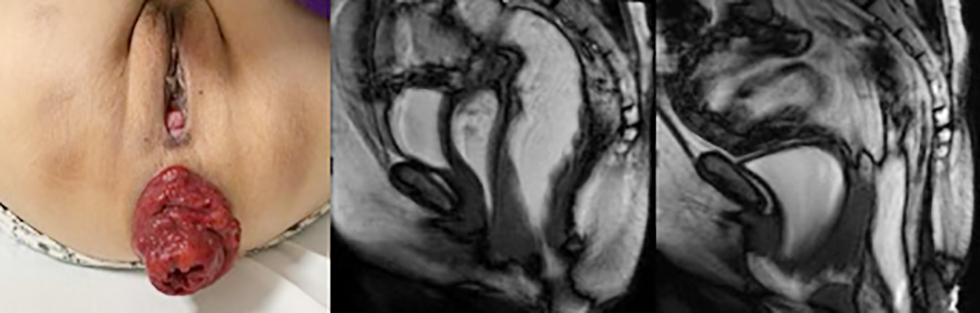
• Preoperative Assessment: Patients underwent clinical examination, medical history review, and specialized investigations, including dynamic pelvic floor MRI and colonoscopy. Standard laboratory tests were performed to evaluate coagulation profile, complete blood count, liver and renal function, and blood glucose. Immuno-logical screening (HIV, HCV, HBsAg), urinalysis, and blood typing were conducted for selection and exclusion based on the established criteria.
• Surgical Intervention: Patients underwent laparoscopic reconstruction of the posterior pelvic floor suspension axis using synthetic mesh with fixation to the sacral promontory.
• Follow-up: Postoperative follow-up was conducted at 1 month and 6 months via direct clinical examination.
• Data Collection: Preoperative, intraoperative, and postoperative parameters, as well as follow-up data, were recorded using a standardized medical record form.
Statistical Analysis
Data were analyzed using SPSS software (version 20.0). Continuous variables were compared using the paired t-test.
Ethical Considerations
The surgical procedure was approved by a multidisciplinary team (MDT), including the Department of Colorectal and Pelvic Floor Surgery, Radiology, and Anesthesiology, with formal authorization from the Board of Directors of Hanoi Medical University Hospital. All patients provided informed consent for surgery after a detailed explanation of potential risks, intraoperative accidents, and postoperative complications. Patient confidentiality was strictly maintained, and data were used solely for scientific research purposes.
SURGICAL TECHNIQUE
Principle: To reconstruct the posterior pelvic floor suspension axis extending from the perineal body to the sacral promontory, thereby restoring the physiological support and suspension mechanisms of the rectum and the pelvic floor.
Figure 1 - Preoperative clinical and MRI findings
Trocar Placement
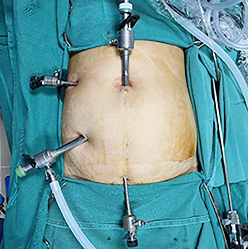
The patient is placed in the supine lithotomy position under general endotracheal anesthesia (fig. 2). Pneumoperitoneum is established, and a four-trocar configuration is utilized:
• A 10-mm umbilical trocar is inserted for the laparoscope.
• A second 10-mm trocar is placed in the right iliac fossa (main working port), approximately 2-3 cm superiorly and medially to the right anterior superior iliac spine (ASIS).
• A 5-mm trocar is inserted in the right flank, at the intersection of the umbilical horizontal line and the right midclavicular line.
• A final 5-mm trocar is placed at the midline (linea alba), just above the pubic symphysis.
Figure 2 - Trocar placement
Rectal Dissection and Exposure of the Perineal Body and Sacral Promontory
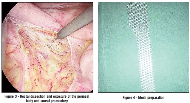
Rectal dissection is performed along the Holy plane (mesorectal plane), initially mobilizing the posterior rectal wall down to the pelvic floor muscles (fig. 3). This maneuver creates a favorable corridor to extend the dissection: anteriorly exposing the rectum down to the perineal body, and right-laterally to the middle rectal vessels. Such comprehensive mobilization is crucial for the uniform suspension of the entire rectum, thereby eliminating the risk of subsequent partial prolapse. Throughout this stage, meticulous attention is paid to preserving the autonomic nerve plexus as well as the middle and inferior right rectal vessels. Subsequently, the sacral promontory is clearly exposed.
Mesh Preparation
A synthetic mesh is tailored to a customized dimension of 3 cm x 15 cm to match the structure of the posterior pelvic floor suspension axis, with its distal portion specifically shaped to match the width of the rectovaginal septum (fig. 4).
Distal Mesh Fixation
The synthetic mesh is introduced into the pelvic cavity (fig. 5). The distal portion of the mesh is securely sutured to the perineal body, and the body of the mesh is fixed to the anterior and right lateral walls of the rectum using non-absorbable braided Ti-Cron 2.0 sutures.
Figure 5 - Distal mesh fixation
Proximal Mesh Fixation and Suspension
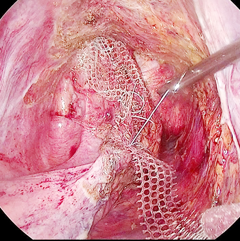
The rectum is suspended cranially without undue tension to ensure a straight anatomical alignment, thereby preventing rectal kinking (angulation), which is a key risk factor for postoperative constipation. To
facilitate this maneuver, a rectal probe (or transanal manipulator) may be inserted into the rectal lumen via the anus to maintain proper axial alignment and ensure optimal tension control (fig. 6). Subsequently, the proximal portion of the synthetic mesh is securely anchored to the sacral promontory using a ProTack device to achieve a stable, complete, and reliable suspension.
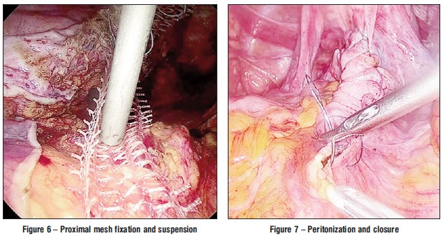
Peritonization and Closure
The pelvic peritoneum is completely re-peritonized, and the Douglas pouch is elevated above the synthetic mesh, which effectively prevents the risk of enterocele or bowel entrapment (fig. 7). Furthermore, this elevation creates a peritoneal scaffold that redistributes and shares the mechanical tension, thereby providing a more sustainable structural support to the posterior pelvic floor suspension axis. Finally, carbon dioxide (CO2) is deflated, and the trocar site incisions are closed in layers according to standard protocol.
RESULTS
Our study included 14 patients with a mean age of 75.5 years (range: 61–87 years). The average parity (number of births) was 3.7, ranging from a minimum of 2 to a maximum of 6 children. The mean Body Mass Index (BMI) was 20.09. The average length of the rectal prolapse segment, measured from the anal verge, was 6.21 cm.
Surgical History:
• 01 patient had a history of pelvic organ prolapse surgery.
• 01 patient had previously undergone surgery for rectal prolapse.
• 01 patient had a history of sigmoid colon resection due to diverticular perforation.
Operative and Postoperative Outcomes:
• Operative time: Range 90–180 minutes (mean: 114.64 minutes).
• Mean intraoperative blood loss: 46 mL.
• Length of hospital stay: 2–5 days.
• Intraoperative accidents: 01 case of rectal seromuscular tear occurred during dissection.
Clinical Progress and Functional Outcomes:
• Most patients experienced postoperative pain with a VAS score of 2–4. At the 1-month follow-up, the majority were pain-free, with only 3/14 patients reporting mild pain (VAS score of 2).
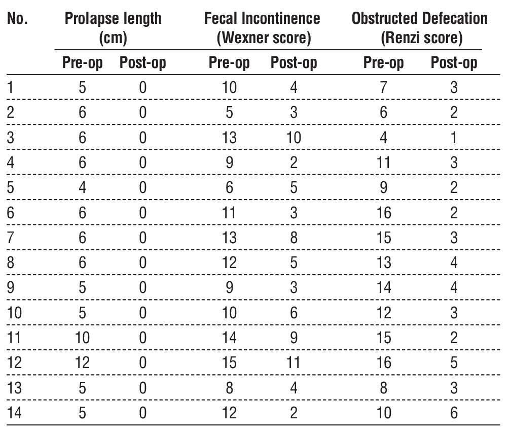
• Patient Quality of Life (QoL) showed significant improvement postoperatively; the mean Agachan - Wexner (Adolfo Renzi) score decreased from 11.14 preoperatively to 3.07 postoperatively.
• The prevalence of fecal incontinence decreased from 85.7% (pre-op) to 28.6% (post-op).
• The prevalence of constipation decreased from 78.8% (pre-op) to 42.9% (post-op).
• No cases of de novo constipation or de novo fecal incontinence were recorded after surgery.
Recurrence: To date, no cases of recurrence have been observed.
DISCUSSION
Our study results showed a mean age of 75.5 years, with the majority of patients having a history of multiparity and a lean body habitus. These characteristics align with the pathogenesis of full-thickness rectal prolapse (FTRP), in which the progressive weakening of the pelvic floor's suspension and support system plays a central role (6,8,9,15,16).
Figure 8 - Clinical appearance before and after laparoscopic PPSA reconstruction
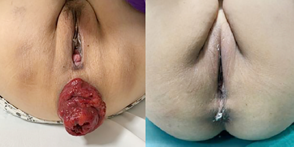
Advanced age is associated with connective tissue degeneration, reduced ligamentous elasticity, and impaired levator ani function (15,18). Furthermore, multiple vaginal deliveries cause cumulative damage to the perineal body and suspensory structures (15,17). These factors do not merely cause instability at a single site but lead to the failure of the entire posterior pelvic floor suspension axis (PPSA), resulting in rectal prolapse. This reinforces the hypothesis that FTRP is not just a rectal pathology but a manifestation of a global pelvic support and suspension disorder (15,16).
Table 1 - Functional outcomes before and after laparoscopic PPSA reconstruction
For decades, the pathogenesis of rectal prolapse was primarily explained by rectal redundancy and mobility (8,10). Based on this view, techniques such as posterior rectopexy and resection-rectopexy were developed to fix the rectum to the sacrum or remove redundant colonic segments (7-9,14). However, recent studies on pelvic floor function indicate that FTRP is closely linked to multi-structural support dysfunction involving the perineal body, levator ani muscles, and pelvic ligamentous systems (15-18). FTRP is increasingly viewed as a systemic pathology rather than an isolated rectal lesion (15,16).
Accordingly, the PPSA reconstruction technique in our study was designed to reconstruct the physiological suspension system between the perineal body, rectum, and sacral promontory, rather than solely repositioning the rectum (1-3,15,16).
Comparison with Other Surgical Techniques
Compared to Posterior Rectopexy
Classically described by Cutait and further developed by various authors, this remains one of the most common techniques for FTRP (7-9). It involves extensive posterior rectal dissection and sacral fixation. Although it achieves low recurrence rates, several studies (including recent meta-analyses) report de novoor worsening constipation in 15–50% of case (7-9).
The primary causes are attributed to:
1. Damage to the autonomic nerve plexus.
2. Loss of physiological rectal mobility (7,8).
In contrast, our technique performs selective posterior dissection - not for fixation, but to facilitate anterior dissection and rectal mobilization. This allows the elevation of the entire rectum, pouch of Douglas, and pelvic floor along a continuous axis while preserving nerve function (11-13,15). This distinction explains why constipation improved rather than increased in our study.
Compared to Ventral Mesh Rectopexy (VMR)
Popularized by André D'Hoore, VMR is considered a significant advancement due to its limited posterior dissection, which reduces nerve damage and post-operative constipation (11). Studies have shown VMR is effective in improving fecal incontinence with low rates of de novo constipation (11-13).
However, some authors note that this method:
1. Does not extend dissection to the perineal body.
2. Only reconstructs a portion of the suspension system.
Consequently, in complex pelvic floor disorders - particularly in elderly women - the outcomes may be incomplete (13,15,16). Our PPSA reconstruction not only preserves nerves like VMR but also extends the reconstruction down to the perineal body, forming a complete suspension axis (15-17).
Compared to Resection-Rectopexy
This is primarily indicated for patients with severe constipation and a redundant sigmoid colon. While it improves constipation via colonic resection, it carries risks associated with anastomosis, such as leaks and infectious complications (7,14). Furthermore, like posterior rectopexy, it fails to address the core issue of pelvic suspension weakening (15,16). Our PPSA technique requires no bowel resection, reducing invasiveness while improving constipation through a physiological mechanism rather than mechanical intervention.
Core Factors of Functional Improvement
The functional improvements recorded in our study can be explained by three core factors:
-
Continuous Suspension Axis: PPSA reconstruction creates a continuous axis from the perineal body to the sacral promontory, rather than a localized anchor point. This ensures even
force distribution across the support system,
preventing force concentration at a single site and maintaining the physiological mobility of the rectum (15,16).
2. Integration of the Perineal Body: The perineal body serves as both the inferior anchor of the rectum and the focal point for various pelvic floor muscles and ligaments (15,17). By integrating the perineal body into the suspension axis, we restore the continuity of the system, improving the coordination between the rectum, anal canal, and levator ani during defecation (15,18).
3. Nerve-Sparing Approach: By minimizing intervention in the posterior and lateral rectal spaces, we preserve the pelvic autonomic plexus, which is essential for regulating motility and defecatory reflexes (11-13). This preservation likely explains the absence of de novo constipation in our cohort (7-9).
The synergy of these three factors - reconstructing a continuous axis, including the perineal body, and preserving nerve function - creates a physiological approach that restores both anatomical position and the global function of the pelvic support system (15,16). This provides a robust basis for the simultaneous improvement of fecal incontinence, constipation, and overall quality of life (13,19,20).
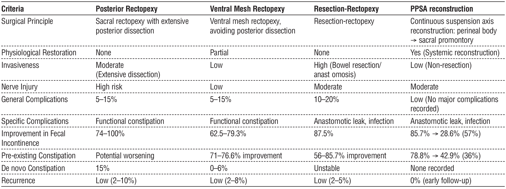
Regarding constipation, posterior rectopexy has been documented to carry a risk of exacerbating this condition, with a de novo constipation rate of approximately 15% and worsening of pre-existing symptoms in up to 50% of patients. This is primarily due to extensive posterior and lateral dissection, which damages the autonomic nerve plexus (7-9). In contrast, ventral mesh rectopexy (VMR) limits de novo constipation (approximately 0–6%) and can improve pre-existing symptoms in 71–76.6% of cases by avoiding posterior dissection (11-13). Resection-rectopexy shows improvement rates ranging from 56% to 85.7%, mainly due to the excision of the redundant sigmoid colon; however, this mechanism is mechanical and may not fully resolve dysmotility if the underlying cause is related to the support system (7,14). In our study, the constipation rate decreased from 78.8% to 42.9%, demonstrating significant improvement without any recorded cases of de novo constipation, suggesting the benefits of reconstructing a physiological suspension axis combined with nerve preservation.
For fecal incontinence, all rectopexy techniques show substantial efficacy. Posterior rectopexy improves incontinence in 74-100% of cases, VMR achieves 62.5-79.3%, and resection-rectopexy stands at approximately 87.5% (9,11-14). In our series, the fecal incontinence rate dropped from 85.7% to 28.6% (equivalent to a 57% improvement). While this improvement is comparable to existing methods, the distinction lies in the mechanism: PPSA reconstruction not only restores anatomical position but also re-establishes coordination between the rectum, anal canal, and pelvic floor through a continuous suspension axis that includes the perineal body (15-17).
Regarding accidents and complications, posterior rectopexy carries a risk of autonomic nerve damage due to extensive dissection, while resection-rectopexy poses additional risks associated with anastomosis (leaks, infections), with overall complication rates of 10–20% (7-9,14). VMR has an overall complication rate of 5–15%, with mesh-related complications accounting for 1–3% (12,22). In our study, we recorded one case of rectal seromuscular tear (7.1%), which was identified and repaired intraoperatively without leading to postoperative complications. No other major complications occurred, with a mean intraoperative blood loss of 46 mL. These results indicate that the PPSA reconstruction technique is safe and acceptable, with accidents being primarily localized and manageable.
Regarding recurrence rates, traditional rectopexy methods all achieve low rates, ranging from 2% to 10% depending on the technique and follow-up duration. In our series, no cases of recurrence (0%) have been recorded during the early follow-up period. This outcome may be related to the reconstruction of a continuous suspension axis from the perineal body to the sacral promontory, which promotes even force distribution rather than concentration at a single fixation point, thereby increasing the durability of the support system (15,16).
Table 2 - Comparison of surgical techniques for full-thickness rectal prolapse
In our study, the Adolfo Renzi score decreased from 11.14 preoperatively to 3.07 postoperatively, indicating a marked improvement in defecatory function and quality of life. This scoring system is a comprehensive assessment tool reflecting factors related to bowel control and its impact on daily activities (19). Therefore, the significant score reduction reflects not just individual symptom changes but a comprehensive functional recovery of the anorectal system.
In summary, while traditional techniques rely primarily on the principle of rectal fixation with certain functional trade-offs (particularly constipation), the PPSA reconstruction technique allows for simultaneous improvement across multiple criteria, including reduced constipation, improved fecal incontinence, a low complication rate, and no early recurrence. These findings support the hypothesis that an approach based on reconstructing the physiological suspension system, rather than simple organ fixation, can play a vital role in optimizing outcomes for full-thickness rectal prolapse (15,16).
CONCLUSION
Our study demonstrates that laparoscopic reconstruction of the posterior pelvic floor suspension axis is a feasible, safe, and functionally effective method for treating full-thickness rectal prolapse. Unlike traditional techniques that focus primarily on rectal fixation, this approach aims to reconstruct a continuous suspension axis, thereby restoring the physiological support mechanism of the pelvic floor.
The significant improvements in fecal incontinence, constipation, and quality of life in this study suggest that direct intervention in the pathogenesis may yield more comprehensive results than purely anatomical corrections. Specifically, the role of the perineal body and force distribution within the entire support system should be viewed as central factors in treatment strategies.
Although these preliminary results are limited by a small sample size and short follow-up period, this technique opens a new direction in the treatment of rectal prolapse, shifting from organ-focused surgery to functional system reconstruction. Controlled studies with larger sample sizes and long-term follow-up are necessary to confirm the superiority of this method and define its place in clinical practice and future treatment guidelines.
Authors’ Contributions
All authors made substantial contributions to one or more critical aspects of the study (conceptualization and design; data acquisition, analysis, or interpretation; drafting the manuscript or critical scholarly revision), approved the final version, and agree to be accountable for all aspects of the work. All authors confirm that they meet the ICMJE authorship criteria.
Acknowledgements
We gratefully thank Hanoi Medical University Hospital and our colleagues at the Department of Coloproctology and Pelvic Floor Surgery for their support throughout the study.
Competing Interests
The authors declare no competing interests.
Funding
This research received no external funding.
Consent to Participate
Waived due to the retrospective design and anonymized data.
Data Availability
The datasets used and/or analyzed during the
current study are available from the corresponding author on reasonable request.
REFERENCES
1. Tran DN, Nguyen AT, Nguyen PD, Nguyen AN, Luu DQ, Nguyen VTT et al. Laparoscopic Fixation of the Anterior Perineal Suspension Axis to the Bilateral Pectineal Ligaments for Recurrent Pelvic Organ Prolapse. Journal of Obstetrics, Gynecology and Cancer Research. 2025;10(9):710-719.
2. D?ng TN, Long TB, D?ng LQ, Phan N?, Vinh NT, Âu H?. ?i?u tr? sa sinh d?c b?ng c? ??nh tr?c treo ?áy ch?u tr??c vào d?i ch?u l??c hai bên qua ph?u thu?t n?i soi. TCNCYH. 2024;178(5):112-120.
3. Tran DN, Nguyen AT, Nguyen AN, Trinh VT. Anatomy and Application of Anterior Perineal Suspension Axis in Treatment of Pelvic Organ Prolapse. Journal of Obstetrics, Gynecology and Cancer Research. 2025;10(12):907-916
4. Townsend CM Jr, Beauchamp RD, Evers BM, Mattox KL, eds. Sabiston Textbook of Surgery: The Biological Basis of Modern Surgical Practice. 21st ed. Philadelphia, PA: Elsevier; 2022.
5. Brunicardi FC, Andersen DK, Billiar TR, Dunn DL, Kao LS, Hunter JG, et al, eds. Schwartz’s Principles of Surgery. 11th ed. New York, NY: McGraw-Hill Education; 2019.
6. Bordeianou L, Paquette IM, Johnson E, Holubar SD, Gaertner W, Feingold DL, et al. Clinical practice guidelines for the treatment of rectal prolapse. Dis Colon Rectum. 2017;60(11):1121-1131.
7. Tou S, Brown SR, Malik AI, Nelson RL. Surgery for complete rectal prolapse in adults. Cochrane Database Syst Rev. 2015;(11): CD001758.
8. Madiba TE, Baig MK, Wexner SD. Surgical management of rectal prolapse. Arch Surg. 2005;140(1):63-73.
9. Varma MG, Wang JY, Berian JR, et al. The American Society of Colon and Rectal Surgeons clinical practice guidelines for the management of rectal prolapse. Dis Colon Rectum. 2021;64(9):1113-1128.
10. Felt-Bersma RJF, Cuesta MA. Rectal prolapse, rectal intussusception, and solitary rectal ulcer syndrome. Gastroenterol Clin North Am. 2001;30(1):199-222.
11. D’Hoore A, Cadoni R, Penninckx F. Long-term outcome of laparoscopic ventral rectopexy for total rectal prolapse. Br J Surg. 2004; 91(11):1500-1505.
12. Consten ECJ, van Iersel JJ, Verheijen PM, Broeders IAMJ. Long-term outcome after laparoscopic ventral mesh rectopexy: an observational study of 919 consecutive patients. Ann Surg. 2015;262(5):742-747.
13. Wijffels NA, Cunningham C, Dixon AR, Lindsey I. Laparoscopic ventral rectopexy for external rectal prolapse is associated with improved fecal continence and constipation. Dis Colon Rectum. 2011;54(7):889-895.
14. Frykman HM, Goldberg SM. The surgical treatment of rectal procidentia. Surg Gynecol Obstet. 1969;129(6):1225-1230.
15. DeLancey JOL. Structural support of the urethra as it relates to stress urinary incontinence: the hammock hypothesis. Am J Obstet Gynecol. 1994;170(6):1713-1723.
16. Petros PE. The Female Pelvic Floor: Function, Dysfunction and Management According to the Integral Theory. 3rd ed. Springer; 2010.
17. Richardson AC. The anatomy of the perineal body. Obstet Gynecol Clin North Am. 1998; 25(4):709-722.
18. Shafik A. A new concept of the anatomy of the anal sphincter mechanism and the physiology of defecation. I. Anatomy of the anal sphincter mechanism. Invest Urol. 1975;13(3):175-182.
19. Renzi A, Brillantino A, Di Sarno G, D’Aniello F. Five-item score for obstructed defecation syndrome: study of validation. Surg Innov. 2013;20(2):119-125.
20. Jorge JMN, Wexner SD. Etiology and management of fecal
incontinence. Dis Colon Rectum. 1993;36(1):77-97.
21. Formijne Jonkers HA, Poierrié N, Draaisma WA, Broeders IAMJ, Consten ECJ. Laparoscopic ventral rectopexy for rectal prolapse and symptomatic rectocele: an analysis of 245 consecutive patients. Colorectal Dis. 2013;15(6):695-699.
22. Evans C, Stevenson ARL, Sileri P, Mercer-Jones MA, Dixon V, Cunningham C, et al. A multicenter collaboration to assess the safety of laparoscopic ventral rectopexy. Dis Colon Rectum. 2015;58(8): 799-807.
Full Text Sources:

Abstract:

Views: 38


For Authors
Journal Subscriptions

Jun 2026
Supplements
Instructions for authors
Online submission
Contact
e-ISSN: 2601 - 1700 (online)
ISSN-L: 2559 - 723X
Journal Abbreviation: Surg. Gastroenterol. Oncol.
Surgery, Gastroenterology and Oncology (SGO) is indexed in:
- SCOPUS
- EBSCO
- DOI/Crossref
- Google Scholar
- SCImago
- Harvard Library
- Open Academic Journals Index (OAJI)
Surgery, Gastroenterology and Oncology (SGO) is an open-access, peer-reviewed online journal published by Celsius Publishing House. The journal allows readers to read, download, copy, distribute, print, search, or link to the full text of its articles.
Time to first editorial decision: 25 days
Rejection rate: 61%
CiteScore: 0.2
 IASGO Society News

Meetings and Courses in 2025
Meetings and Courses in 2024
Meetings and Courses in 2023
Meetings and Courses in 2022
Meetings and Courses in 2021
Meetings and Courses in 2020
Meetings and Courses in 2019
Verona expert meeting 2019

Surgery, Gastroenterology and Oncology applies the Creative Commons Attribution Non Commercial (CC BY-NC 4.0) license, which permits readers to copy and redistribute the material in any medium or format, remix, adapt, build upon the published works non-commercially, and license the derivative works on different terms, provided the original material is properly cited and the use is non-commercial. Please see: https://creativecommons.org/licenses/by-nc/4.0/
Publisher’s Note:
The opinions, statements, and data contained in article are solely those of the authors and not of Surgery, Gastroenterology and Oncology journal or the editors. Publisher and the editors disclaim responsibility for any damage resulting from any ideas, instructions, methods, or products referred to in the content.