Surgery, Gastroenterology and Oncology

|
|
Background: Research has shown that identifying retained stones and bile duct injury is difficult. This study aimed to explore the use of laparoscopic ultrasonography (LUS) in detecting common bile duct (CBD) stones and evaluating biliary anatomy during laparoscopic cholecystectomy for patients with gallbladder stones.
Methods: This case series enrolled 77 adult patients of both genders with symptomatic gallbladder stones who were scheduled for laparoscopic cholecystectomy. Patients underwent LUS to detect CBD stones before proceeding with the cholecystectomy. The primary outcome was the incidence of LUS-detected CBD stones and the ability to visualize the CBD clearly during surgery. Secondary outcomes included the length of hospital stay and the requirement for postoperative endoscopic retrograde cholangiopancreatography (ERCP) or magnetic resonance cholangiopancreatography.
Results: The patients' mean age was 44.08±13.32 years, with female predominance (62.34%). Intraoperative LUS detected CBD stones in 12.99% of patients, with a clear view of the CBD in 93.51% of patients. The mean duration of cholecystectomy was 132.2±6.56 min. Postoperative ERCP was done in 7.79% of patients. The mean hospital stay was 1.26±0.88 days. Obstructive jaundice occurred in 2.6% of patients in whom stones were not detected by LUS.
Conclusion: In adult patients undergoing laparoscopic cholecystectomy, intraoperative LUS is a feasible and useful adjunctive technique for evaluating the biliary architecture and identifying CBD stones, with an acceptable safety profile.
Keywords: common bile duct stones, laparoscopic cholecystectomy, laparoscopic ultrasonography
INTRODUCTION
Gallstone disease is a common gastrointestinal disorder worldwide, with a global prevalence of approximately 6% and evidence of rising incidence in recent decades. The prevalence increases progressively with advancing age, reaching nearly 9% among individuals older than 50 years (1).
Laparoscopic cholecystectomy is the preferred treatment modality for symptomatic gallstone disease and is recognized as the gold‑standard approach in current clinical practice (2). Laparoscopic cholecystectomy has proved to be relatively safe when performed by an experienced surgeon, provided that there is clear visualization of the biliary ducts to prevent injury during the operation (3, 4). A key adverse consequence of laparoscopic cholecystectomy is the failure to detect asymptomatic common bile duct (CBD) stones, which may present later as retained or recurrent gallstone disease. Among patients with gallbladder stones, the prevalence of concomitant CBD stones widely ranges from 1 to 15% (5). The identification of CBD stones and the prevention of bile duct damage necessitate meticulous examination and delineation of the biliary tract.
Laparoscopic ultrasonography (LUS) uses an ultrasound transducer that is inserted through laparoscopic ports to scan the bile ducts from the hepatic duct confluence to the ampulla, allowing a comprehensive view of the CBD. It provides real-time, high-resolution visualization of the CBD, enabling the detection of stones and the prevention or prompt identification of biliary duct damage during laparoscopic cholecystectomy (6). The procedure has gained wide popularity due to its minimally invasive nature, cost-effectiveness, and lack of risk of radiation. Previous studies have found LUS comparable to, if not superior to, intraoperative cholangiography (7-9). For CBD stones, studies reported that the sensitivity of LUS ranges from 80% to 96% and specificity ranges from 96% to 100%, which aligns with or exceeds the sensitivity and specificity of intraoperative cholangiography (75% to 87% and 98% to 99%, respectively) (7,10,11).
However, the routine adoption of LUS varies across medical settings, and current evidence on its feasibility and impact on intraoperative decision-making remains limited. This debate is particularly important in patients who are classified as low risk for CBD stones according to the criteria of the American Society for Gastro-enterology (ASGE) (12) and the European Society of Gastrointestinal Endoscopy (ESGE) (13), as current ASGE/ESGE guidelines recommend cholecystectomy with optional intraoperative imaging. However, a small but clinically meaningful proportion of those patients can still have occult CBD stones that become detected either intra- or postoperatively (14,15). Assessment of the efficacy of LUS in detecting CBD stones in this subset of patients may reduce unnecessary ductal exploration, avoid postoperative ERCP, and potentially lower the risk of missed stones without subjecting the entire cohort to ionizing radiation or more costly imaging techniques. Therefore, this study aimed to explore the use of LUS in detecting common bile duct (CBD) stones and evaluating biliary anatomy during laparoscopic cholecystectomy for patients with gallbladder stones.
MATERIAL AND METHODS
Ethical Considerations
The Ethics Committee at Cairo University's Faculty of Medicine approved the study (approval code: MD-191-2022; date: 4/8/2022). All participants provided written informed consent before undergoing the procedure. Confidentiality of patients’ data was strictly maintained by keeping anonymous data collection sheets after assigning a unique identifier to each patient.
Study Design, Setting, and Location
This case series was conducted at Cairo University Hospitals between August 2022 and April 2024.
Eligibility Criteria
Patients of both sexes, aged 18 to 65, with symptomatic gallbladder stones and normal liver enzymes who were scheduled for elective laparoscopic cholecystectomy were included in this study. Exclusion criteria were the detection of a CBD stone on transabdominal ultrasound, serum bilirubin greater than 4 mg/dL, refusal to participate, presence of psychiatric disorders, or presence of disease contraindicating a laparoscopic procedure.
Data Collection
A comprehensive history was obtained from each patient, followed by a meticulous clinical examination and a series of laboratory investigations. These investigations encompassed a complete blood count, liver function tests (alanine aminotransferase, aspartate aminotransferase, total and direct bilirubin, albumin, alkaline phosphatase enzyme, and gamma glutamyl transpeptidase), renal function tests (blood urea and serum creatinine), serum electrolytes, and a coagulation profile.
The diagnosis of gallbladder stones was determined by a thorough review of the patient's medical history, which included a complaint of right hypochondrial pain. Additionally, pelvic-abdominal ultrasonography was performed, which revealed the presence of gallbladder stones.
Patients were risk‑stratified for bile duct stones according to the ASGE criteria. Those with no predictors (normal liver enzymes, bilirubin ≤ 4 mg/dL, and no CBD stone or dilation on ultrasound) were classified as low risk and, per guideline recommendations, proceeded directly to elective laparoscopic cholecystectomy without routine preoperative magnetic resonance cholangiopancreatography (MRCP).
Preoperative MRCP was done for all patients with intermediate risk, such as those with abnormal liver function tests (elevated aminotransferases, alkaline phosphatase enzyme, or total bilirubin up to 4 mg/dL), age older than 55 years, or dilated common bile duct (> 6 mm) on abdominal ultrasonography without a visualized stone. We included in our study only patients with no MRCP-evident CBD stones.
Surgical Procedure
Subsequent to the insertion of the ports for the laparoscopic cholecystectomy, forceps were utilized to grasp and elevate the gallbladder's fundus and the liver's right lobe to enable the visualization of the
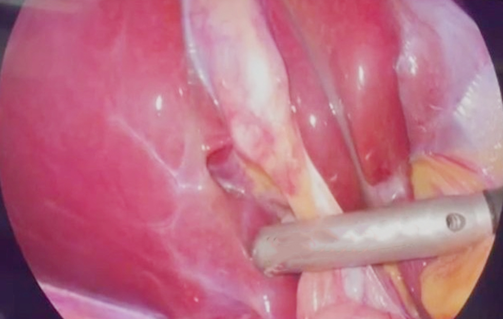
hepatoduodenal ligament. The epigastric route was chosen to facilitate the insertion of the LUS transducer. Subsequently, the transducer was delicately positioned over the hepatoduodenal ligament, thereby enabling the evaluation of the CBD (fig. 1).
Figure 1 - Intraoperative laparoscopic ultrasonography of the common bile duct. The laparoscopic ultrasound probe is positioned over the hepatoduodenal ligament during laparoscopic cholecystectomy to obtain an ultrasound view of the common bile duct. The surrounding hepatobiliary structures are visualized in the operative field.
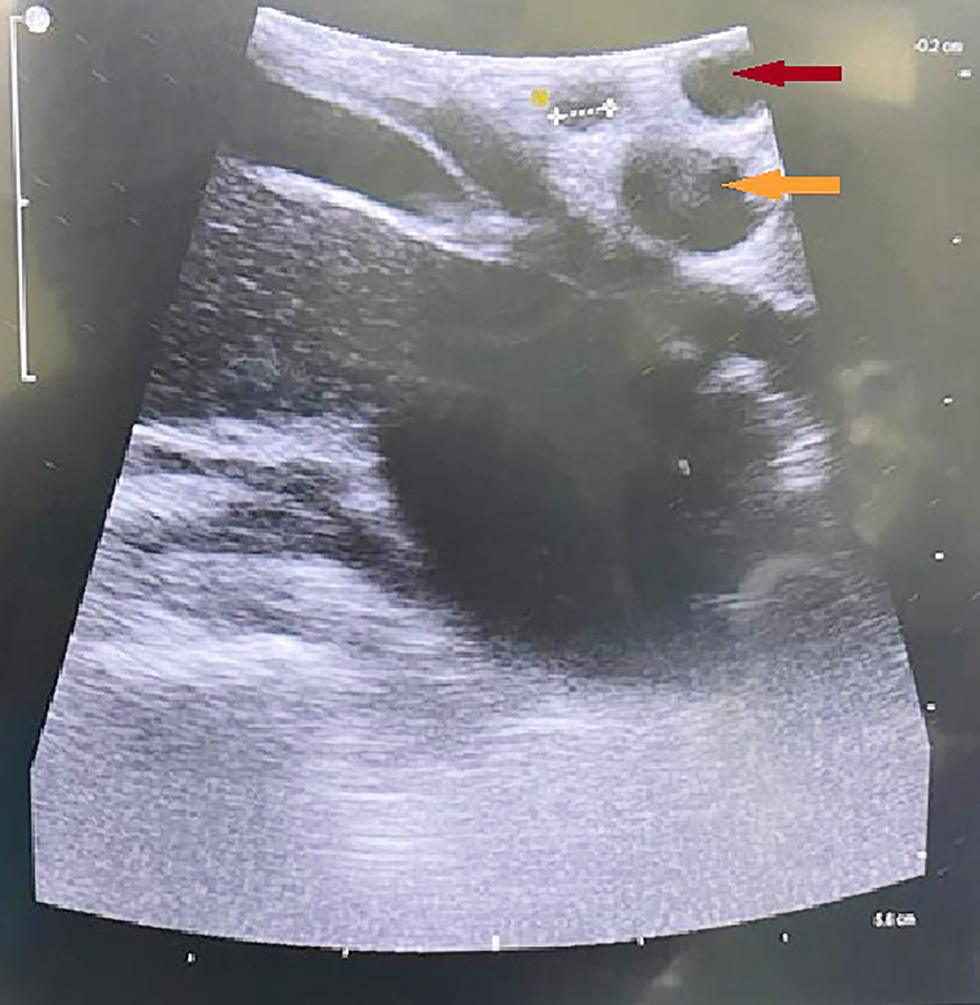
The radiological examination was initiated at the point of intersection between the cystic duct and the common hepatic duct, where the CBD was first observed. Furthermore, the CBD was tracked all the way down to the duodenum. The portal vein is located posteriorly and has been likened to the iconic figure of "Mickey's head," while the hepatic artery and the CBD constitute the anteriorly positioned right and left "ears," respectively. Using this "Mickey Mouse" appearance or sign facilitated identification of the hepatoduodenal ligament anatomy and measurement of the CBD diameter (fig. 2).
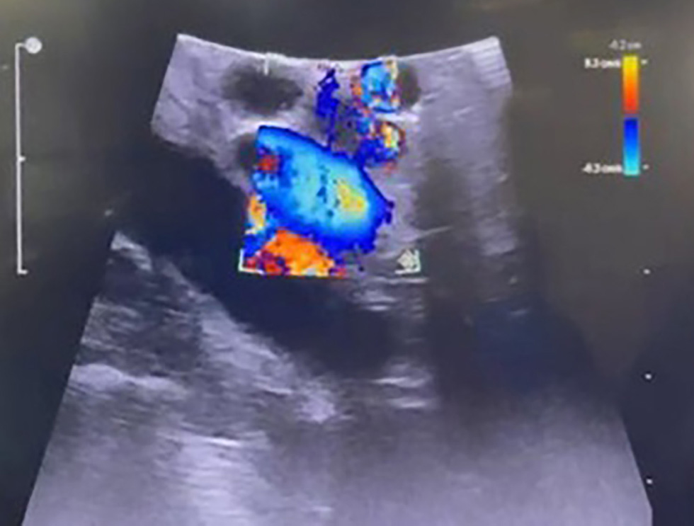
The portal vein and hepatic artery (which exhibit flow) were readily distinguished from the CBD, which lacks internal flow, by employing the Doppler function (fig. 3).
Figure 3 - Doppler function to distinguish the portal vein and hepatic artery from the common bile duct. The portal vein and hepatic artery (which exhibit flow, blue coloration) were readily distinguished from the common bile duct (yellow arrow), which lacks internal flow.
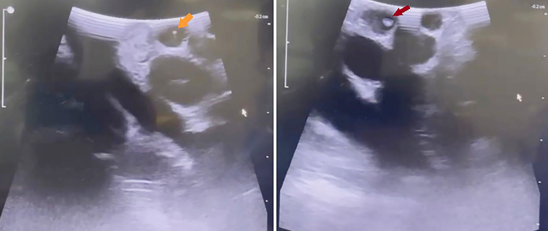
While some small CBD stones can theoretically pass spontaneously, small CBD stones < 4 mm (fig. 4 a) were cleared and managed through a transcystic route (16, 17) that minimizes invasiveness during definitive cholecystectomy to avoid future complications like cholangitis, pancreatitis, or jaundice. Transcystic CBD exploration provides a single-stage laparoscopic option for managing choledocholithiasis without requiring a formal choledochotomy. The required instrumentation includes mechanical balloon dilatation catheters (4-8 mm) for cystic duct expansion, Fogarty-type biliary balloon catheters (4–6 Fr) for stone extraction, and laparoscopic clips or Hem-o-lok clips for secure cystic duct stump closure. The cystic duct is identified, skeletonized, and milked toward the gallbladder to clear small debris. A distal clip is placed at the gallbladder-cystic duct junction to prevent continued stone spillage. A partial transverse incision is made on the anterolateral aspect of the cystic duct using laparoscopic micro-scissors or a hook cautery, taking care not to completely transect the duct. If the cystic duct lumen is narrow or spastic, step-wise mechanical dilatation is performed using a dedicated balloon dilator pressurized with saline. A balloon catheter is inflated proximal to the stone and swept distally to push the stone through the sphincter of Oddi into the duodenum, often augmented by intravenous glucagon (1 mg) or smooth muscle relaxants to relax the ampullary sphincter (Balloon Flushing / Push Technique). Complete lithic clearance (removal or passage) of all identified CBD stones without retained fragments was confirmed by intraoperative LUS using a high-frequency flexible laparoscopic ultrasound probe applied directly along the porta hepatis and hepatoduodenal ligament, demonstrating an anechoic CBD lumen without hyperechoic foci or posterior acoustic shadowing.
Meanwhile, larger stones ≥4 mm (fig. 4 b) were cleared by postoperative endoscopic retrograde cholangiopancreatography (ERCP) in the same hospital admission as a standard alternative for laparoscopic CBD exploration.
Figure 4 - (a) Small stone (orange arrow) < 4 mm in size detected in the common bile duct.
(b) Large stone (red arrow) > 4 mm in size detected in the common bile duct.
Postoperative Complications
A meticulous observation of immediate postoperative complications was conducted, including anesthesia-related risks (such as cardiovascular complications) as well as procedure-related risks (such as pain, infection, bleeding, and leakage). The assessment of complications was conducted in accordance with the Clavien-Dindo classification system, a comprehensive framework utilized to categorize postoperative complications (18).
Study Outcomes
The primary outcomes were the incidence of LUS-detected CBD stones and the ability to visualize the CBD clearly during surgery. Secondary outcomes included the length of the procedure, the length of hospital stay, and the requirement for ERCP or magnetic resonance cholangiopancreatography (MRCP).
Regarding the detection of stones, CBD stones identified by intraoperative LUS were recorded. Following surgery, all patients were clinically monitored for evidence suggestive of retained CBD stones, including the development of jaundice and/or abnormal liver function tests. When such findings occurred, further diagnostic evaluation by ERCP was performed, and the presence of LUS-missed stones was confirmed. Patients who did not develop clinical or biochemical evidence suggestive of retained CBD stones during postoperative follow-up were considered not to have clinically apparent retained CBD stones. According to the management protocol for such cases at our institution, ERCP was not performed systematically in all patients but was undertaken selectively when clinically indicated.
Statistical Analysis
The statistical analysis was conducted using the Statistical Package for the Social Sciences (SPSS v26, IBM, Inc., Chicago, IL, USA). Quantitative data were presented as the mean and standard deviation, median, and range, while qualitative data were presented as numbers and percentages. Statistical significance was adopted at p-value ˂ 0.05.
RESULTS
As illustrated in table 1, the mean age of the included patients was 44.08 ± 13.32 years. Most patients were female (62.34%), had no history of abdominal surgeries (66.23%), and were free of comorbidities (79.22%).
Table 1 - Baseline characteristics, previous abdominal operations, and comorbidities (n=77)
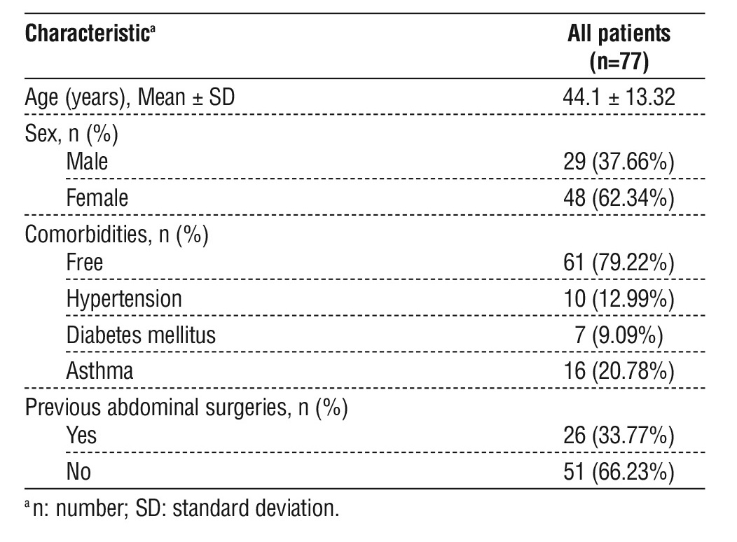
Table 2 demonstrates that patients exhibited hemoglobin levels ranging from 10.1 to 13.4 g/dL. The values of total leukocyte count, platelet count, hepatic aminotransferases, bilirubin, serum albumin, alkaline phosphatase, gamma glutamyl transpeptidase, amylase, lipase, and C-reactive protein were within normal ranges. Preoperative ultrasonography revealed that the CBD diameter ranged from 2 to 9 mm, with a mean of 3.64 ± 1.22 mm. Furthermore, 5.19% of patients had intrahepatic biliary dilatation (IHBRD), and 7.79% had a history of pancreatitis before surgery. In this study, four patients (5.19%) with intermediate-risk predictors underwent preoperative MRCP, which was negative for CBD stones.
Table 2 - Preoperative laboratory and ultrasonographic investigations (n = 77)

As shown in table 3, the operating time ranged from 122 to 151 minutes. Intraoperative LUS exhibited a clear view of the CBD in the majority of patients (93.51%), with detection of CBD stones in 12.99% of patients. The average stone size was 5.7 ± 1.9 mm. None of the patients developed intraoperative bleeding or injury to the CBD. No CBD injuries or hemorrhagic complications were observed. Postoperative jaundice occurred in two (2.6%) patients (Clavien–Dindo Grade IIIb), with no reported cases of leakage. Postoperative ERCP was performed in six (7.79%) patients: four patients in whom CBD stones were identified by LUS, and two other patients who had LUS-undetected CBD stones and experienced post-operative biliary obstruction. Thus, CBD stones were identified in 12 of the 77 patients (15.58%) based on intraoperative LUS findings and selective postoperative ERCP. No life-threatening complications (Grade IV) or postoperative mortalities (Grade V) were recorded. The mean duration of hospitalization was 1.26 ± 0.88 days.
Table 3 - Operative time, intra-operative laparoscopic ultrasonography, and postoperative follow-up (n = 77)

DISCUSSION
The present study aimed to investigate the efficacy of LUS in detecting CBD stones and evaluating biliary anatomy during laparoscopic cholecystectomy in patients with gallbladder stones. The study population comprised patients with symptomatic gallstone disease who had normal preoperative biochemical findings and no identified CBD stones on preoperative investigations. According to the 2019 ASGE criteria, patients without additional predictors of CBD stones are considered low risk, whereas patients older than 55 years or those with a dilated CBD (>6 mm) are classified in the intermediate-risk category. Thus, the cohort included patients spanning the low- and intermediate-risk spectrum. The ASGE guidelines allow for cholecystectomy without additional ductal imaging in low-risk patients while recommending endoscopic ultrasonography, MRCP, IOC, or intraoperative LUS for intermediate-risk patients (12). This provided a clinically relevant population in which to evaluate whether intraoperative LUS could detect occult CBD stones despite reassuring preoperative biochemical and imaging findings.
In the current case series, baseline patient characteristics agreed with studies conducted in several
countries, where adult females were more predominant than males (19,20).
The overall procedural time in our case series was 132.22 ± 6.56 minutes, including the laparoscopic cholecystectomy approach, intraoperative LUS, and the subsequent therapeutic steps. The time required specifically for LUS was not recorded separately in our cohort; therefore, the individual contribution of LUS to the total operative time cannot be quantified. The total procedure duration can be influenced by the learning curve associated with the adoption of LUS and the overall technique. Accordingly, the reported operative time should be interpreted in the context of the study setting and operator experience and cannot be generalized to other settings or regarded as the definitive time required for the technique. Previous systematic reviews found that the mean imaging time for LUS ranged between approximately 5 and 10 minutes (11,21). Future studies should assess the learning curve of LUS imaging time and subsequent therapeutic steps to allow a more precise assessment of its contribution to overall operative duration.
In the current study, intraoperative LUS detected CBD stones in 12.99% of patients, and a clear CBD
visualization was achieved in 93.51% of cases, enabling reliable stone detection. The literature reports that intraoperative LUS can enable clear visualization of the CBD in approximately 85 to 98% of patients undergoing laparoscopic cholecystectomy (11,21). As regards the detection of CBD stones, a systematic review found that studies reported sensitivities for LUS ranging between 80 and 100%, with the reported specificities ranging between 96 and 100% (11). The detection rate in our case series should be cautiously interpreted as exploratory analyses, as gold standard tests were not consistently performed in all included patients, precluding a formal assessment of diagnostic accuracy. Despite the exploratory nature of the analysis, the present study contributes preliminary data on CBD stone detection in a low- to intermediate-risk population that has not been adequately characterized in previous research. As LUS remains inherently operator-dependent, achieving optimal CBD visualization demands specialized training and practiced expertise in ultrasound imaging. Future studies should assess the impact of surgeon expertise and learning curve on achieving CBD visualization rates and the diagnostic accuracy of LUS for detecting CBD stones.
In the present study, obstructive jaundice occurred in 2.59% of patients postoperatively, while no patient experienced leakage. The absence of intraoperative bleeding or CBD injury suggests intraoperative LUS safety. However, the incidence of bile duct injuries following laparoscopic cholecystectomy ranges from 0.3 to 0.5% (3,19), requiring a much larger sample size to assess whether the use of LUS reduces the incidence of these relatively uncommon adverse events.
Limitations
This single-center study with a relatively small sample size may yield findings less generalizable to other institutions. This is particularly important in interpreting the absence of rare complications, as the possibility of the occurrence of such rare events in larger cohorts cannot be ruled out. In addition, the study design, lacking a comparator and a consistent gold standard test in all cases, did not allow for formal assessment of the diagnostic performance of LUS in detecting occult CBD stones.
Conclusions
In adult patients who underwent laparoscopic cholecystectomy, LUS was a feasible and useful adjunctive method for detecting CBD stones and evaluating the biliary anatomy. However, future studies with larger sample sizes are required to confirm the safety of LUS and its potential incremental value in reducing the incidence of bile duct injury during laparoscopic cholecystectomy. Future trials should include a gold standard test for assessing the diagnostic accuracy of LUS in detecting CBD stones. In addition, the studies should assess the potential impact of operator and surgeon experience as well as the stage of the learning curve on the success of LUS in achieving clear visualization of CBD and detecting stones.
Conflicts of Interest: None.
Source of Funding: None.
REFERENCES
1. Ning Q, Liu F, Fang Y, Zhu X, Liu J, Li Z. Estimating global prevalence of gallbladder stones in general population from 2000 to 2024: systematic review and meta-analysis. Ann Med. 2025;57(1): 2570795.
2. Zhang Y, Dai X, Duan R, Wei L. Research progress in the treatment of gallstones with laparoscopic and endoscopic surgery: a narrative review. BMC Surg. 2025;25(1):238.
3. Symeonidis S, Mantzoros I, Anestiadou E, Ioannidis O, Christidis P, Bitsianis S, et al. Biliary Anatomy Visualization and Surgeon Satisfaction Using Standard Cholangiography versus Indocyanine Green Fluorescent Cholangiography during Elective Laparoscopic Cholecystectomy: A Randomized Controlled Trial. J Clin Med. 2024; 13(3):864.
4. Gupta V, Jain G. Safe laparoscopic cholecystectomy: Adoption of universal culture of safety in cholecystectomy. World J Gastrointest Surg. 2019;11(2):62-84.
5. McNicoll CF, Pastorino A, Farooq U, Froehlich MJ, Hill CRS. Choledocholithiasis. In: StatPearls. Treasure Island (FL): StatPearls Publishing. 2026 Jan. 2023 Jul 10.
6. de'Angelis N, Catena F, Memeo R, Coccolini F, Martínez-Pérez A, Romeo OM, et al. 2020 WSES guidelines for the detection and management of bile duct injury during cholecystectomy. World J Emerg Surg. 2021;16(1):30.
7. Jamal KN, Smith H, Ratnasingham K, Siddiqui MR, McLachlan G, Belgaumkar AP. Meta-analysis of the diagnostic accuracy of laparoscopic ultrasonography and intraoperative cholangiography in detection of common bile duct stones. Ann R Coll Surg Engl. 2016; 98(4):244-9.
8. Kumar SK, Shehata DG, Cetrulo LN, Ignacio R, Chiu J, Davis BR, et al. SAGES guidelines for the use of intraoperative imaging of the common bile duct. Surg Endosc. 2025;39(11):7091-7102.
9. Ball E, Waters N, Cooper N, Talati C, Mallick R, Rabas S, et al. Evidence-Based Guideline on Laparoscopy in Pregnancy: Commissioned by the British Society for Gynaecological Endoscopy (BSGE) Endorsed by the Royal College of Obstetricians & Gynaecologists (RCOG). Facts Views Vis Obgyn. 2019;11(1):5-25.
10. Dili A, Bertrand C. Laparoscopic ultrasonography as an alternative to intraoperative cholangiography during laparoscopic cholecystectomy. World J Gastroenterol. 2017;23(29):5438-5450.
11. Awan B, Elsaigh M, Marzouk M, Sohail A, Elkomos BE, Asqalan A, et al. A Systematic Review of Laparoscopic Ultrasonography During Laparoscopic Cholecystectomy. Cureus. 2023;15(12):e51192.
12. Buxbaum JL, Abbas Fehmi SM, Sultan S, Fishman DS, Qumseya BJ, Cortessis VK, et al. ASGE guideline on the role of endoscopy in the evaluation and management of choledocholithiasis. Gastrointest Endosc. 2019;89(6):1075-105.e15.
13. Manes G, Paspatis G, Aabakken L, Anderloni A, Arvanitakis M, Ah-Soune P, et al. Endoscopic management of common bile duct stones: European Society of Gastrointestinal Endoscopy (ESGE) guideline. Endoscopy. 2019;51(5):472-91.
14. Varone A, Mendoza A, Gelbard R, Tracy B, Poulose B. Suspected common bile duct stones: Which test is best? Surg Endosc. 2025; 39(10):6525-6529.
15. Wang X, Li Z. Management of common bile duct stones: a comprehensive review. Front Surg. 2025;12:1658784.
16. Tsuda S. Choledocholithiasis: The Society of American Gastrointestinal and Endoscopic Surgeons; ND [Available from: https://www.sages.org/wiki/choledocholithiasis/.
17. Haggerty S, Richardson W, Santos B, Fanelli R, Brunt LM, Stefanidis D. Clinical Spotlight Review: Laparoscopic Common Bile Duct Exploration: The Society of American Gastrointestinal and Endoscopic Surgeons; ND [Available from: https://www.sages.org/ publications/guidelines/clinical-spotlight-review-laparoscopic-common-bile-duct-exploration/.
18. Clavien PA, Barkun J, de Oliveira ML, Vauthey JN, Dindo D, Schulick RD, et al. The Clavien-Dindo Classification of Surgical Complications: Five-Year Experience. Ann Surg. 2009;250(2): 187-96.
19. Song Y, Ma Y, Xie FC, Jin C, Yang XB, Yang X, et al. Age, gender,
geographic and clinical differences for gallstones in China: a nationwide study. Ann Transl Med. 2022;10(13):735.
20. Rodriguez Gatta D, Huidobro L, Petermann-Rocha F, Van de Wyngard V, Godoy F, Cid V, et al. Sex disparities in gallstone disease: insights from the MAUCO prospective population-based cohort study. BMJ Open Gastroenterol. 2024;11(1):e001457.
21. Edebo A, Andersson J, Gustavsson J, Jivegård L, Ribokas D, Svanberg T, et al. Benefits and risks of using laparoscopic ultrasonography versus intraoperative cholangiography during laparoscopic cholecystectomy for gallstone disease: a systematic review and meta-analysis. Surg Endosc. 2024;38(9):5096-5107.
Full Text Sources:

Abstract:

Views: 400


For Authors
Journal Subscriptions

Jun 2026
Supplements
Instructions for authors
Online submission
Contact
e-ISSN: 2601 - 1700 (online)
ISSN-L: 2559 - 723X
Journal Abbreviation: Surg. Gastroenterol. Oncol.
Surgery, Gastroenterology and Oncology (SGO) is indexed in:
- SCOPUS
- EBSCO
- DOI/Crossref
- Google Scholar
- SCImago
- Harvard Library
- Open Academic Journals Index (OAJI)
Surgery, Gastroenterology and Oncology (SGO) is an open-access, peer-reviewed online journal published by Celsius Publishing House. The journal allows readers to read, download, copy, distribute, print, search, or link to the full text of its articles.
Time to first editorial decision: 25 days
Rejection rate: 61%
CiteScore: 0.2
 IASGO Society News
Meetings and Courses in 2025
Meetings and Courses in 2024
Meetings and Courses in 2023
Meetings and Courses in 2022
Meetings and Courses in 2021
Meetings and Courses in 2020
Meetings and Courses in 2019
Verona expert meeting 2019

Surgery, Gastroenterology and Oncology applies the Creative Commons Attribution Non Commercial (CC BY-NC 4.0) license, which permits readers to copy and redistribute the material in any medium or format, remix, adapt, build upon the published works non-commercially, and license the derivative works on different terms, provided the original material is properly cited and the use is non-commercial. Please see: https://creativecommons.org/licenses/by-nc/4.0/
Publisher’s Note:
The opinions, statements, and data contained in article are solely those of the authors and not of Surgery, Gastroenterology and Oncology journal or the editors. Publisher and the editors disclaim responsibility for any damage resulting from any ideas, instructions, methods, or products referred to in the content.