Surgery, Gastroenterology and Oncology

|
|
Background: Entamoeba gingivalis is a non-pathogenic ameba that inhabits the human oral cavity and occasionally other sites. Entamoeba gingivalis may become pathogenic in cancer patients under chemotherapy. The aim of this study is to investigation of Entamoeba gingivalis in cancer patients compared to healthy individuals (as a control), in the Thi-Qar Province of Iraq.
Methods: Samples were collected from individuals with diseased periodontal sites and from individuals with healthy periodontal sites from cancer patients and control groups in a hospital Al-Haboubi Teaching Hospital for the period from August 2023 to December 2023, for detection of E. gingivalis. Samples were diagnosed by microscopic staining with Giemsa stain, and the recovered positive isolates were subjected to molecular analysis.
Results: Amplification of DNA extracted from E. gingivalis of both cancer patients and healthy people using 18 rDNA revealed PCR products with 204 pb size for both diseased and healthy people. This study indicated that there are significant differences (p<0.05) in the percentage of E. gingivalis infections among cancer patients with periodontitis these have a higher infection rate (62%) than those who without (56 %), as well as control group was 56 % in individuals with periodontitis, and 30 % in individuals without. The current study showed the overall rate of infection of E. gingivalis by PCR technique was) 66.13%.
Conclusion: This study showed the sequence was identical and there was no difference in sequence between all isolates.
Introduction
Entamoeba gingivalis is a protozoan microorganism that is mostly found in the human oral cavity, and it has also been detected in the genitourinary tract, Transmission occurs through contaminated food or oral equipment, mouth droplets, and kissing (1). E. gingivalis has a cosmopolitan distribution with a worldwide prevalence of 37% (2). Some studies proposed an association between E. gingivalis and periodontal diseases (2,3).
Periodontal disease is an inflammatory condition affecting the gingiva, due to localized inflammation of the periodontium on exposure to dental plaque (4); Dental plaque build-up results in gingivitis, though reversible when oral hygiene measures are introduced. However, if left untreated, it may involve the underlying periodontal tissue (5). Cancer is a serious problem affecting the health of all human societies (6), it is a state in which cell proliferation occurs abnormally when it becomes uncontrollable and able to invade other cells. Additionally, the number of cancer patients has been clearly rising in recent years (7), Chronic infection or toxins production, immune evasion, and immuno-logical suppression are all important mechanisms that can lead to cancer (8). This study was designed to investigate microscopic and molecular of Entamoeba gingivalis in oral cavity of cancer patients with periodontitis and compare with healthy oral persons.
Materials and Methods
Collection of Samples
A total of 300 swab samples were collected: 100 each from cancer patients with diseased and healthy periodontal sites who visited the Thi-Qar Oncology center at Al-Haboubi teaching hospital and received chemotherapy. Furthermore 100 swab samples were collected from healthy individuals, with 50 having diseased periodontal sites and 50 with healthy periodontal sites, as a control group. During the period from August 2023 to December 2023.
Microscopic Examination
Samples were applied directly onto slides and stabilized using methanol. Then, they were stained with giemsa stain for a duration of 15 to 20 minutes and examined by microscope under (100X).
Molecular Examination
Genomic DNA Extraction
Genomic DNA was extracted from oral wash samples (saliva and dental plaque) by using gSYNCTM DNA Extraction Kit (Geneaid Taiwan) done according to company instruction. The PCR primers for detection Entamoeba gingivalis based on small subunit ribosomal RNA gene, The primers used, SSU (F-AGGAATGAACGGAACGTACA) and rDNA (R-CCATTTCCTTCTTCTATTGTTTCAC) (9).
PCR Reaction Compounds
The compounds that used for PCR reaction were master mix (13 µl), primer forward (1 µl), primer reverse (1 µl), DNA template ((30-100ng)) (4 µl), and free water (6 µl), totally give volume of 25 µl.
Protocol of PCR Amplification
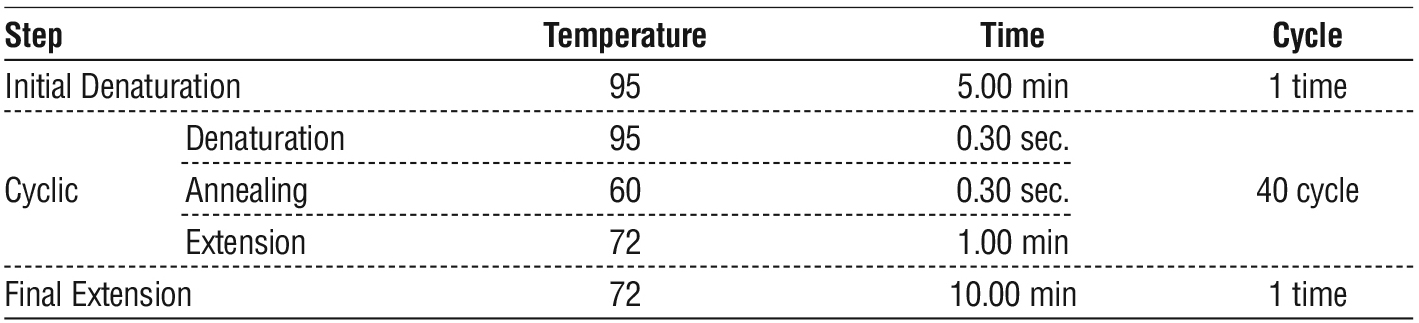
The protocol used in this study is shown in a table 1.
Table 1 - Protocol of PCR amplification.
Sequencing and Alignment of Genes
The PCR products amplified with two primers were sequenced by Macrogen Company (South Korea). NCBI BLAST tool was used to check the results of this program, and to detect any DNA alterations in the sequences of the gene.
Statistical Analysis
The data of the present study were statistically analysis by using SPSS (Statistical Package of Social Science version 26) based in using both descriptive and non-parametric Chi-Square at p. value < 0.05 (10).
Results
Microscopic Examination
The current results demonstrated that cancer patients with unhealthy periodontal sites had a higher prevalence of E. gingivalis compared to cancer patients with healthy periodontal sites, and the rate of infection was 62% and 34%, respectively. As well as control group was 56 % in individuals with diseased periodontal sites, and 30 % in individuals with healthy periodontal sites, as shown in table 2.
Table 2 - Percentage of infection with Entamoeba gingivalis in cancer patients and control group by microscopic examination.

Molecular Examination
Forty-eight samples were taken from positive microscopic examination, only 40 (83.33%) were positive after PCR examination, as well as 14 samples from negative microscopic examination, only 1(7.14%) were positive after by pcr examination. The overall rate of infection of E. gingivalis by PCR technique (figs. 1, 2) was 66.13%, as shown in table 3.
Table 3 - The percentage of Entamoeba gingivalis infection among cancer patients based on type of diagnostic method.
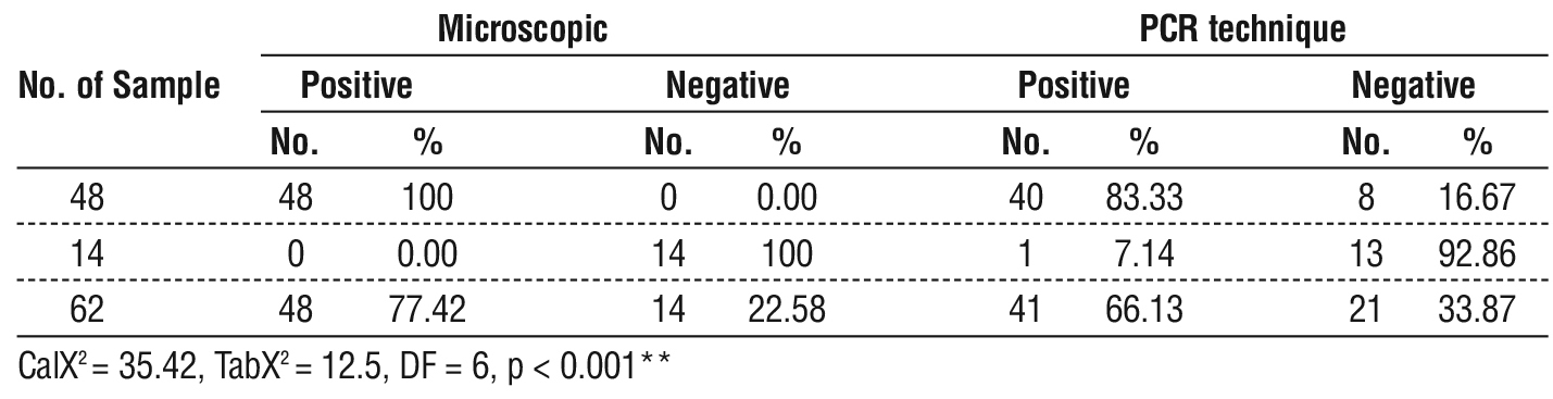
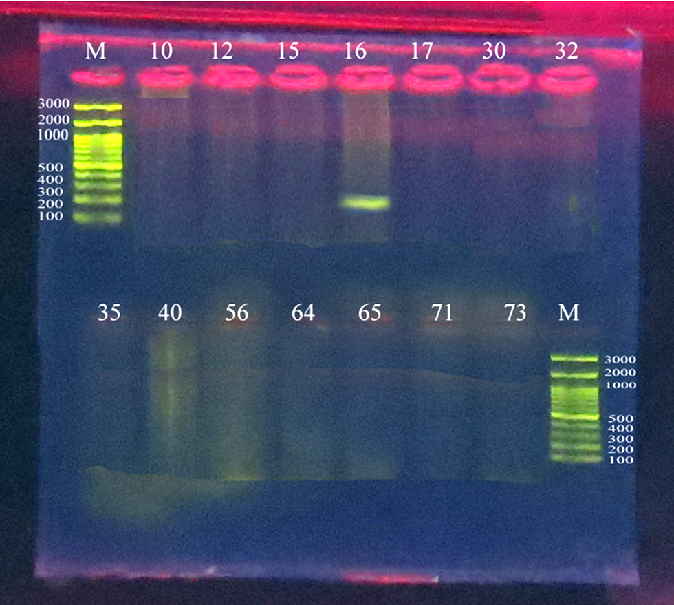
Figure 1 - Electrophoresis of a PCR product of Entamoeba gingivalis with a size of 204 bp, using agarose gel at a concentration of 1.5%, at a voltage of 70 V and 85 mA, for 45 minutes. For positive microscopy.
The sequence was identical and there was no difference in sequence between all isolates (each of the cancer patients with periodontitis, cancer patients without periodontitis, control with periodontitis and control without periodontitis). In this study: The isolate No.1 (Accession No. = LC804957; size = 204bp) identified 100% with specie Accession No. = OR405086. The isolate No.2 (Accession No. = LC804968; size= 204bp) identified 100% with specie Accession No. = OR405085. The isolate No.3 (Accession No. = LC804969; size = 204bp) identified 100% with specie Accession No. = OQ225457. The isolate No.4 (Accession No. = LC804970; size= 204bp) identified 100% with specie Accession No. = PP335301. The isolate No.50 (Accession No. = LC804958; size= 204bp) identified 100% with specie Accession No. = OR405085. The isolate No.72 (Accession No. = LC804964; size= 204bp) identified 100% with specie Accession No. = PP335301. The isolate No.74 (Accession No. = LC804962; size= 204bp) identified 100% with specie Accession No. = OR405085.
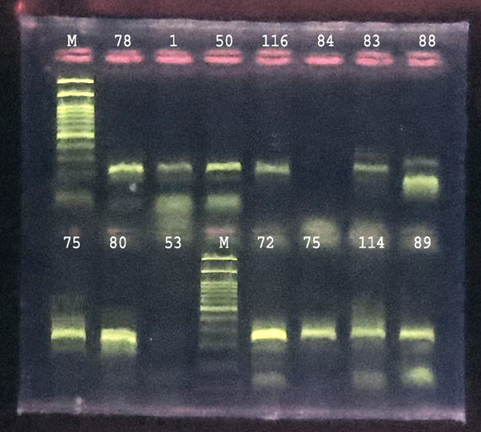
Figure 2 - Electrophoresis of a PCR product of Entamoeba gingivalis with a size of 204 bp, using agarose gel at a concentration of 1.5%, at a voltage of 70 V and 85 mA, for 45 minutes. For negative microscopy.
 The isolate No.75 (Accession No. = LC804965; size= 204bp) identified 100% with specie Accession No. = KX061779. The isolate No.78 (Accession No. = LC804956; size= 204bp) identified 100% with specie Accession No. = KX061779. The isolate No.80 (Accession No. = LC804963; size= 204bp) identified 100% with specie Accession No. = OQ225457. The isolate No.83 (Accession No. = LC804960; size= 204bp) identified 100% with specie Accession No. = PP335301. The isolate No.88 (Accession No. = LC804961; size= 204bp) identified 100% with specie Accession No. = KX061779. The isolate No.89 (Accession No. = LC804967; size= 204bp) identified 100% with specie Accession No. = KX061779. The isolate No. 114 (Accession No. = LC804966; size= 204bp) identified 100% with specie Accession No. = KX061779. The isolate No. 116 (Accession No. = LC804959; size= 204bp) identified 100% with specie Accession No. = OQ225457, (figs. 3, 4).
 Sequencing
Figure 3 - The Phylogenic tree of Entamoeba gingivalis Isolates between our isolates & reference copies in NCBI

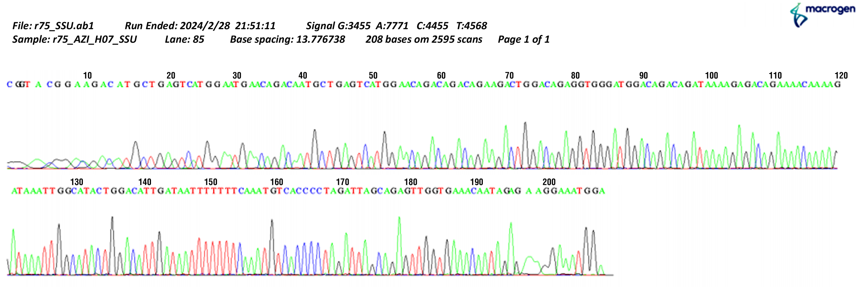
Figure 4 - The sequence analysis of 18S rRNA Gene "Entamoeba gingivalis" by "Korean Macrogen company"
Discussion
Entamoeba gingivalis is one of the members of the wide range of oral resident pathogens in humans, particularly found in dental plaques, surfaces of gingiva or teeth, interdental spaces and carious lesions (2). The results of current study revealed that individuals with diseased periodontitis sites had a higher prevalence of E. gingivalis than individuals without periodontitis, in both groups (cancer patients and control), this result is consistent with (11) who found that the rate was (77%) of inflamed periodontal sites and (22%) of healthy sites, (12) where was found that 73.84% of inflamed periodontal sites and 50% of healthy periodontal sites exhibited this phenomenon, additionally, maybe the rate of E. gingivalis infection increases in individuals with periodontitis due to changes in the oral environment and the accumulation of bacteria, which makes it more suitable for the growth and reproduction of E. gingivalis. One of the most important causes of periodontitis is poor of oral hygiene, which is the same reason for increased parasite infection. Therefore, we notice that the rate of E. gingivalis infection increases in individuals with periodontitis.
The results of this study shows the results of PCR technique, where the overall rate of infection of E. gingivalis was (66.13%) and this result is consistent with (13,14), where the percentages were (76%) and (64%), respectively, the study did not consistent with (15,16) where the percentages were (11.4 %) and (16 %), respectively. PCR is more sensitive technique than direct microscopy of culture, but the use of both PCR and culture method is suggested for environmental water samples to gain more complete results of the real presence of amoeba (17).
The sequence was identical and there was no difference in sequence between all isolates, this study did not consistent with (18) who found fifty percent of the samples were found positive for the E. gingivalis ST1 subtype, (24%) were found positive for the E. gingivalis ST2 kamaktlii variant. The reason may be attributed to the shortness of the studied region of the DNA strand (204 bp).
Conclusion
This study investigated the Entamoeba gingivalis in cancer patients compared to healthy individuals (as a control), in the Thi-Qar Province of Iraq. This study showed the sequence was identical and there was no difference in sequence between all isolates.
Conflicts of Interest
The authors declare no conflict of interest.
Informed Consent Statement
The informed consent was obtained.
References
1. Örsten S, Sahin C, Y?lmaz E, Akyön Y. First molecular detection of Entamoeba gingivalis subtypes in individuals from Turkey. Pathog Dis. 2023;81:ftad017.
2. Badri M, Olfatifar M, Abdoli A, Houshmand E, Zarabadipour M, Abadi PA, et al. Current global status and the epidemiology of Entamoeba gingivalis in humans: A systematic review and meta-analysis. Acta Parasitol. 2021;66(4):1102–13.
3. Garcia G, Ramos F, Maldonado J, Fernandez A, Yáñez J, Hernandez L, et al. Prevalence of two Entamoeba gingivalis ST1 and ST2-kamaktli subtypes in the human oral cavity under various conditions. Parasitol Res. 2018;117(9):2941–8.
4. Savage A, Eaton KA, Moles DR, Needleman I. A systematic review of definitions of periodontitis and methods that have been used to
identify this disease. J Clin Periodontol. 2009;36(6):458-67.
5. Oladokun AO, Ogboru P, Opeodu OI, Lawal AO, Falade MO. Prevalence of Entamoeba gingivalis and Trichomonas tenax among patients with periodontal disease attending Dental Clinic, University College Hospital, Ibadan. Trop Parasitol. 2023; 13(2):107–13.
6. Al-Mugdadi SFH. Diagnostic Biomarkers Related to Cancer Detection and Treatment: A Review Article. University of Thi-Qar. J. Sci. 2021;8:3–6.
7. Hameed F, Khalaf A. Serological detection for Entamoeba histolytica among cancer patients suffering from diarrhea in Thi-Qar province/ Southern Iraq. J. Sci. 2022;9(2):3–6.
8. Hamza Abbas H, Abdulrazzaq Gati Al nasery M, Abdulhussein jarullah B. Molecular and Phylogenic study of some Gastrointestinal Bacteria and Viruses associated with Cancer in the South area of Iraq. J. Sci. 2022;9(1):9–18.
9. Hussian RS. Molecular detection of Entamoeba gingivalis using polymerase chain reaction. Pakistan Journal of Biotechnology. 2017;14(3):351–4.
10. Krishnaiah PR. A Hand Book of Statistics. Vol. 1. Motilal Banarsidass Publishe; 1980.
11. Bao X, Wiehe R, Dommisch H, Schaefer AS. Entamoeba gingivalis causes oral inflammation and tissue destruction. J Dent Res. 2020;99(5):561–7.
12. Nuaimi BN, Al-Taee AF, AL-Kattan MM. The effect of Entomoeba gingivalis infection in patients with diabetes and high pressure on the level of Malonaldehyde and Glutathione. Int J Health Sci (IJHS) . 2022;6143–52.
13. Sh R, Sabitha R, Shyam S, Arunmozhi S, Kadhiresan U, Govindarajan S. Incidence of the oral protozoa - Entamoeba gingivalis in a hospitalbased population in South India - A preliminary study. Journal of Oral Disease Markers. 2018;2(1):1–4.
14. Vlasa A, Bud A, Lazar L, Lazar AP, Herbert A, Bud E. Association of Entamoeba gingivalis with Periodontal Disease-Systematic Review and Meta-Analysis. Medicina (Kaunas). 2024;60(5):736.
15. Sharifi M, Jahanimoghadam F, Babaei Z, Mohammadi MA, Sharifi F, Hatami N, et al. Prevalence and associated-factors for Entamoeba gingivalis in adolescents in southeastern Iran by culture and PCR, 2017. Iran J Public Health. 2020;49(2):351–9.
16. Rahdar M, Abolfazli-Karizi S, Pedram H. The comparison of Entamoeba gingivalis presence in healthy and periodontitis patients by using direct examination and PCR methods. Jundishapur J Health Sci. 2019; In Press.
17. Muslim, Al-Badran A. Morphological and Molecular Identification of Free-living Amoebae Acanthamoeba spp. Isolated from Environmental and clinical Sources in Thi-Qar province / Iraq. Annals of the Romanian Society for Cell Biology. 2021;25:6704–14.
18. Zaffino M, Dubar M, Debourgogne A, Bisson C, Machouart M. Development of a new TaqMan PCR assay for the detection of both Entamoeba gingivalis genotypes. Diagn Microbiol Infect Dis. 2019;95(4):114886.
Full Text Sources:
Abstract:
Views: 2686

For Authors
Journal Subscriptions

Dec 2025
Supplements
Instructions for authors
Online submission
Contact
e-ISSN: 2601 - 1700 (online)
ISSN-L: 2559 - 723X
Journal Abbreviation: Surg. Gastroenterol. Oncol.
Surgery, Gastroenterology and Oncology (SGO) is indexed in:
- SCOPUS
- EBSCO
- DOI/Crossref
- Google Scholar
- SCImago
- Harvard Library
- Open Academic Journals Index (OAJI)
Surgery, Gastroenterology and Oncology (SGO) is an open-access, peer-reviewed online journal published by Celsius Publishing House. The journal allows readers to read, download, copy, distribute, print, search, or link to the full text of its articles.
Time to first editorial decision: 25 days
Rejection rate: 61%
CiteScore: 0.2

Meetings and Courses in 2025
Meetings and Courses in 2024
Meetings and Courses in 2023
Meetings and Courses in 2022
Meetings and Courses in 2021
Meetings and Courses in 2020
Meetings and Courses in 2019
Verona expert meeting 2019

Surgery, Gastroenterology and Oncology applies the Creative Commons Attribution Non Commercial (CC BY-NC 4.0) license, which permits readers to copy and redistribute the material in any medium or format, remix, adapt, build upon the published works non-commercially, and license the derivative works on different terms, provided the original material is properly cited and the use is non-commercial. Please see: https://creativecommons.org/licenses/by-nc/4.0/
Publisher’s Note:
The opinions, statements, and data contained in article are solely those of the authors and not of Surgery, Gastroenterology and Oncology journal or the editors. Publisher and the editors disclaim responsibility for any damage resulting from any ideas, instructions, methods, or products referred to in the content.