Surgery, Gastroenterology and Oncology
|
|
Abbreviations: GBV: Gallbladder Volvulus; CT: Computed Tomography; CRP: C-reactive Protein.
Introduction: Gallbladder volvulus (GBV) is a rare but potentially fatal condition, often clinically and radiologically indistinguishable from acute cholecystitis. Prompt diagnosis and surgical management are crucial to prevent severe complications.
Case Presentation: We report the case of an 85-year-old male admitted with right upper quadrant pain and imaging findings suggestive of acute cholecystitis. Abdominal ultrasonography and CT scan revealed a markedly distended gallbladder with pericholecystic fluid but without definitive signs of torsion. Laparoscopic exploration revealed a 360° counterclockwise torsion of the gallbladder around its mesentery, confirming the diagnosis of gallbladder volvulus. The patient underwent successful detorsion and laparoscopic cholecystectomy. The postoperative course was uneventful, and histopathology confirmed acute gangrenous cholecystitis.
Discussion: The clinical presentation of GBV often mimics acute cholecystitis, making preoperative diagnosis particularly challenging. Imaging findings, although non-specific, can retrospectively suggest the diagnosis, particularly when a floating and horizontally positioned gallbladder is observed.
Conclusion: This case underscores the importance of considering GBV in the differential diagnosis of atypical or severe acute cholecystitis presentations, especially in elderly patients. Increased clinical awareness is critical to achieving early surgical intervention and improving patient outcomes.
Keywords: gallbladder volvulus, acute cholecystitis, laparoscopic cholecystectomy
INTRODUCTION
Gallbladder volvulus (GBV), also known as gallbladder torsion, is a rare pathological condition that often mimics acute cholecystitis. First described by Wendel in 1898 (1), fewer than 500 cases have been documented in the English medical literature (2). GBV involves the rotation of the gallbladder on its mesentery, occluding the cystic duct and cystic artery, which can lead to ischemia, necrosis, and life-threatening complications such as perforation or sepsis (3). Although rare, with an occurrence rate of approximately 1 in 365,520 hospitalizations (2), GBV carries a mortality rate of up to 6% if left untreated (3).
Most cases occur in elderly patients, particularly women aged between 65 and 75 years, and are often associated with anatomical variations such as a ‘floating gallbladder’ (4). Symptoms, including right upper quadrant pain and imaging findings of gallbladder wall thickening or biliary sludge, frequently overlap with acute cholecystitis, complicating diagnosis. Only about 25% of cases are diagnosed preoperatively, with most being identified intraoperatively, leading to delays in critical intervention (5).
Despite advances in diagnostic tools, the lack of distinguishing features makes the preoperative diagnosis of GBV particularly challenging. Heightened clinical suspicion and timely surgical management are crucial, as delayed cholecystectomy significantly increases morbidity and mortality risks (3).
This article presents a case report of a male patient who was initially diagnosed with acute cholecystitis, in whom GBV was identified intraoperatively.
This case highlights the significant diagnostic challenges posed by GBV and emphasizes the critical role of prompt surgical intervention in improving patient outcomes.
CASE PRESENTATION
An 85-year-old male was admitted to the emergency department with a three-day history of abdominal pain localized to the right upper quadrant.
He reported associated nausea but denied vomiting, jaundice, or fever. His medical history included a previous gastrectomy for a perforated gastric ulcer more than 20 years ago, and bilateral inguinal hernioplasty performed via an anterior approach.
His chronic medications included cyanocobalamin, pyridoxine, thiamine (0.2 mg + 200 mg + 100 mg), and lorazepam 2.5 mg.
On physical examination, there was tenderness upon palpation of the epigastrium and right hypochondrium, with a positive Murphy’s sign. Laboratory analysis revealed leukocytosis of 17,200/µL and elevated C-reactive protein (CRP) at 151 mg/L, without other significant abnormalities.
Abdominal ultrasonography (fig. 1) showed a liver with regular contours and normal size and shape. The hepatic parenchyma was homogenous, without nodular lesions. Intrahepatic and extrahepatic bile ducts were of normal caliber. The gallbladder appeared markedly distended (5.6 × 13.4 cm), with pronounced wall thickening and trabeculation. Perivesicular fluid with septations was noted, though no organized collections or wall discontinuity were evident. The lumen contained biliary sludge and small echogenic foci suggestive of microlithiasis. These findings were consistent with acute or sub-acute cholecystitis.
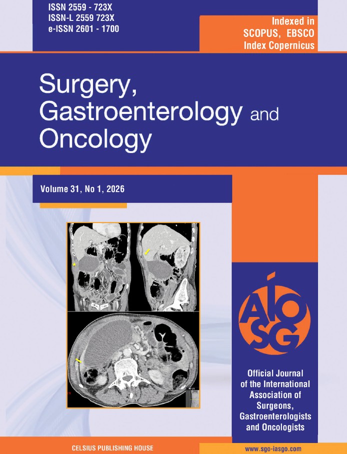
Subsequent computed tomography (CT) of the abdomen (fig. 2) confirmed significant gallbladder distension, further supporting the diagnosis of acute cholecystitis.
The patient was then scheduled for laparoscopic cholecystectomy.
Intraoperatively, the gallbladder appeared ischemic and markedly distended, with a counterclockwise torsion of approximately 360° around its mesentery, findings consistent with gallbladder volvulus (fig. 3).
After performing detorsion of the gallbladder, the procedure proceeded as a standard cholecystectomy, with careful dissection of Calot’s triangle and ligation of the cystic duct and artery. The gallbladder was removed through the umbilical port using an endobag (fig. 4), and a surgical drain was placed.
The postoperative course was uneventful, and the patient was discharged on the third postoperative day following removal of the abdominal drain.
The anatomopathological report revealed findings consistent with acute gangrenous cholecystitis.
DISCUSSION
Gallbladder volvulus (GBV) is an exceptionally rare entity among abdominal emergencies, characterized by torsion of the gallbladder around its mesenteric axis (1).
While torsion is more commonly associated with hollow organs such as the sigmoid colon or cecum, its
Figure 1 - Ultrasound images of the gallbladder demonstrating signs consistent with acute gallbladder pathology. Findings include gallbladder distentsion, wall thickening, intraluminal sludge or debris, and pericholecystic fluid - features that may suggest acute cholecystitis or,

in rare cases, G
Figure 2 - CT images: (a) distended lumen of the gallbladder (star); (b, c): the distended lumen of the gallbladder transitions to the narrow

fulcrum points at its neck (yellow arrow) occurrence in the gallbladder is uncommon and often under-recognized (2).
The rarity of this condition, combined with its non-specific clinical presentation, poses significant challenges in both diagnosis and management (3).
GBV can present as either clockwise or counterclockwise torsion, with the degree of rotation ranging from incomplete (<180°) to complete torsion (>180°) <<)>>.
Gallstones are found in only 24–32% of cases (5), emphasizing the importance of considering GBV as a distinct diagnostic entity, particularly in elderly patients without a history of gallstones.
The etiology of GBV remains unclear, although several anatomical and physiological factors appear to contribute. A redundant mesentery, allowing excessive gallbladder mobility, is the most recognized predisposing factor (6). This anatomical variation, present in approximately 4% of the population, may involve the entire gallbladder and cystic duct, or only the cystic duct itself (7). Age-related changes, such as <> of the surrounding tissues and the reduction of visceral fat, further exacerbate organ mobility and predispose older
Figure 3 - Intraoperative image showing a markedly distended and ischemic gallbladder. A complete counterclockwise torsion around the mesentery is visible, consistent with gallbladder volvulus

Figure 4 - Surgical specimen: markedly distended gallbladder with a longitudinal diameter of approximately 15 cm, presenting

signs of ischemia and areas of necrosis individuals to this condition (8). Additional risk factors include rapid weight loss, hepatic atrophy, and vigorous peristaltic activity of adjacent organs (9).
Consequently, GBV predominantly affects older adults, with a peak incidence between 60 and 80 years of age (10).
Clinically, GBV mimics acute cholecystitis, presenting with right upper quadrant pain, nausea, vomiting, and fever (11).
On physical examination, patients may exhibit tenderness in the right hypochondrium, a palpable mass, or peritoneal signs (12).
Laboratory findings typically reveal elevated inflammatory markers, although abnormalities in liver function tests are less common (3).
The overlap of these features with acute cholecystitis often delays the diagnosis of GBV, resulting in the majority of cases being identified intraoperatively (5).
The diagnostic challenge lies in the non-specificity of imaging findings. Ultrasound and CT may demonstrate a distended, thick-walled gallbladder, often without gallstones (6).
MRI, though less frequently utilized, can be particularly helpful in identifying complications such as necrosis or intramural hemorrhage (13).
Kitagawa et al. proposed specific imaging criteria for GBV, including fluid collection, a horizontally positioned floating gallbladder, and signs of ischemia or inflammation (7).
Although these proposed imaging criteria appear promising, they still require further validation in clinical practice.
In fact, in our case, several of these features were present. On ultrasonography (fig. 1), there was a clear presence of pericholecystic fluid completely surrounding the gallbladder.
Furthermore, on axial CT images (fig. 2 c), the gallbladder appeared horizontally positioned and “floating,” deviating from its usual anatomical relationship with the liver bed.
These findings, although subtle, retrospectively correlate well with the imaging criteria suggested by Kitagawa et al. and could have raised suspicion for gallbladder volvulus preoperatively.
Prompt surgical intervention is critical for GBV to prevent complications such as perforation and peritonitis, which can lead to <> and increased mortality (8).
Emergency cholecystectomy, either laparoscopic or open, remains the definitive treatment (9).
Laparoscopy, in particular, has gained traction as a safe and effective approach, offering advantages such as reduced postoperative morbidity and faster recovery (14).
The procedure is facilitated by the detorsion of the gallbladder and its careful dissection to prevent iatrogenic injury (6).
Early intervention results in excellent outcomes, while delayed treatment is associated with a mortality rate of up to 5% (3).
CONCLUSION
Gallbladder volvulus (GBV) is a rare and potentially life-threatening condition that is often misdiagnosed as acute cholecystitis due to overlapping clinical and imaging features.
Preoperative diagnosis remains challenging, with most cases being identified intraoperatively.
Prompt recognition and timely laparoscopic cholecystectomy are essential to prevent severe complications such as ischemia and necrosis.
This case highlights the importance of considering GBV in atypical presentations of acute cholecystitis, particularly in elderly patients.
Increased clinical awareness and more frequent reporting are crucial to improving diagnostic accuracy and patient outcomes
Conflicts of Interest
The authors declare no conflicts of interest.
Source of Funding
No funding was received for the preparation of this article.
REFERENCES
1. Wendel AV. A case of floating gallbladder and kidney complicated by torsion of the gallbladder. Ann Surg. 1898;27(2):199-202.
2. Reilly DJ, Kalogeropoulos G, Thiruchelvam D. Torsion of the gallbladder: a systematic review. HPB (Oxford). 2012;14(10):669-672.
3. Gross RE. Volvulus of the gallbladder. Am J Dis Child. 1938;56(1): 152-154
4. Losanoff JE, Kjossev KT. Complete torsion of the gallbladder. J Am Coll Surg. 1999;189(1):100-101.
5. Kitagawa H, Nakada K, Enami T, Yamaguchi T, Kawaguchi F, Nakada M, et al. Two cases of torsion of the gallbladder diagnosed preoperatively. J Pediatr Surg. 1997;32(11):1567-1569.
6. Pu TW, Fu CY, Lu HE, Cheng WT, Cheng YJ. Gallbladder torsion: A case report and review of the literature. World J Gastroenterol. 2010; 16(4):410-413.
7. Akashi M, Tanaka H, Narimatsu H, Yoshida K, Wada H, Matsumoto S, et al. Torsion of the gallbladder: Report of a case and review of the literature. Surg Today. 2001;31(5):459–463.
8. García-León R, González-Sánchez AJ, Martín-Lorenzo JG, Téllez- Molina MJ, Torralba-Martínez JA, Minguela-Puras A, et al. Vólvulo de vesícula biliar: retos diagnósticos y quirúrgicos. Cir Cir. 2016; 84(2):153–158.
9. Chou CF, Mak CW, Lai HC, Chang JM. Gallbladder torsion detected using computed tomography and magnetic resonance cholangiopancreatography: A case report. J Radiol Case Rep. 2012;6(3): 26-33
10. Boonjun J, Charoensak A, Apisarnthanarak P. Gallbladder torsion: An unexpected intraoperative finding. Clin Med Res. 2018;16(1-2):38- 40.
11. Lau WY, Fan ST, Wong SH. Acute torsion of the gallbladder in the aged: A re-emphasis on clinical diagnosis. Aust N Z J Surg. 1982; 52(5):492-494.
12. Lavoie J, Gregoire R. Torsion of the gallbladder. Can J Surg. 1983; 26(4):339-340.
13. Nakao A, Matsuda T, Funabiki S, Mori T, Koguchi K, Iwado T, et al. Gallbladder torsion: case report and review of 245 cases reported in the Japanese literature. J Hepatobiliary Pancreat Surg. 1999;6(4): 418-21.
14. Ravikumar R, Thomas JS. Torsion of the gallbladder: a systematic review and case report. Ann R Coll Surg Engl. 2010;92(6): W23-W26.
Full Text Sources:

Abstract:

Views: 425


For Authors
Journal Subscriptions

Mar 2026
Supplements
Instructions for authors
Online submission
Contact
e-ISSN: 2601 - 1700 (online)
ISSN-L: 2559 - 723X
Journal Abbreviation: Surg. Gastroenterol. Oncol.
Surgery, Gastroenterology and Oncology (SGO) is indexed in:
- SCOPUS
- EBSCO
- DOI/Crossref
- Google Scholar
- SCImago
- Harvard Library
- Open Academic Journals Index (OAJI)
Surgery, Gastroenterology and Oncology (SGO) is an open-access, peer-reviewed online journal published by Celsius Publishing House. The journal allows readers to read, download, copy, distribute, print, search, or link to the full text of its articles.
Time to first editorial decision: 25 days
Rejection rate: 61%
CiteScore: 0.2
 IASGO Society News
Meetings and Courses in 2025
Meetings and Courses in 2024
Meetings and Courses in 2023
Meetings and Courses in 2022
Meetings and Courses in 2021
Meetings and Courses in 2020
Meetings and Courses in 2019
Verona expert meeting 2019

Surgery, Gastroenterology and Oncology applies the Creative Commons Attribution Non Commercial (CC BY-NC 4.0) license, which permits readers to copy and redistribute the material in any medium or format, remix, adapt, build upon the published works non-commercially, and license the derivative works on different terms, provided the original material is properly cited and the use is non-commercial. Please see: https://creativecommons.org/licenses/by-nc/4.0/
Publisher’s Note:
The opinions, statements, and data contained in article are solely those of the authors and not of Surgery, Gastroenterology and Oncology journal or the editors. Publisher and the editors disclaim responsibility for any damage resulting from any ideas, instructions, methods, or products referred to in the content.